Mass Spectrometry Based Protein Sequencing

Mass Spectrometry Based Protein Sequencing

Service Details

Microscopic 3D render of a protein structure with intricate folds and molecular bonds in a deep navy and teal clinical atmosphere.
Protein Sequencing Service

Mass Spectrometry Based Protein Sequencing

Creative Proteomics supports protein, peptide, and antibody characterization with terminal sequencing, de novo analysis, peptide mapping, intact mass measurement, and site-specific PTM identification.

Terminal Sequencing De Novo Analysis Peptide Mapping PTM Characterization

Service Scope

Sequencing workflows for known, unknown, and modified proteins

biotech

Terminal Sequencing

Determine N-terminal and C-terminal sequence information for structural characterization.

De Novo Sequencing

Reconstruct unknown protein or antibody sequences without prior database records.

Peptide Mapping

Verify sequence coverage, key peptide regions, and molecular consistency.

PTM Analysis

Detect and localize phosphorylation and other post-translational modifications.

Proteins

Support for purified proteins, gel bands, and complex research samples.

Peptides

Peptide-level evidence for sequence confirmation and site localization.

Antibodies

Sequence reconstruction and characterization for challenging antibody projects.

Why Choose Mass Spectrometry for Protein Sequencing?

Mass spectrometry based protein sequencing analysis is the analysis of the amino acid sequence of a sample (protein, antibody, peptide). Mass spectrometry for peptide and protein sequence determination includes bottom-up proteomics and top-down proteomics.

The former is the mainstream technology of mass spectrometry sequencing, i.e., the protein is cleaved into small fragments by specific enzymatic or chemical hydrolysis, and then the molecular weight of each product peptide is detected by mass spectrometry, and the obtained peptide spectrum data is entered into a database to search for known proteins corresponding to it to obtain the protein sequence to be measured.

This method can effectively identify amino acid isomers and post-translational modifications. Due to the introduction of cleavage, it is difficult to resolve and grasp the overall sequence information of the protein using this method, and it is difficult to identify proteins with low content.

The latter method starts from the whole protein molecule, which can be introduced into the mass spectrometry by electrospray or matrix-assisted laser-resolved ionization, and then dissociated and analyzed by tandem mass spectrometry.

Diagram illustrating bottom-up and top-down protein sequencing methodologies using mass spectrometry.

Mass Spectrometry vs. Edman Degradation

Mass spectrometry and Edman degradation are both used for protein sequence analysis, but they differ significantly in sample compatibility, sequence depth, and modification analysis. For modern protein sequencing projects, mass spectrometry offers broader analytical coverage and greater flexibility for complex samples.

Feature Mass Spectrometry Edman Degradation
N-terminal blocking or modification Sequence analysis can still be performed through peptide-level or intact-protein workflows. Blocked N-termini may prevent sequencing.
Sample purity requirement Can handle purified samples and more complex mixtures. Typically requires very high purity.
PTM analysis Supports phosphorylation, glycosylation, and other PTM characterization. Limited for comprehensive PTM analysis.
Sequence coverage Broad coverage through bottom-up and top-down approaches. Usually limited to N-terminal residues.
Complex sample compatibility Suitable for mixed or difficult samples. Less suitable for complex mixtures.
Sensitivity Effective for low-abundance targets with modern high-resolution instruments. More challenging when sample amount is limited.
Leu/Ile differentiation May require specialized fragmentation or orthogonal strategies. Can be distinguished directly.

Mass spectrometry is especially valuable for complex mixtures, modified proteins, monoclonal antibodies, and projects requiring broader sequence coverage or structural characterization.

Comprehensive Protein Sequencing Services

At Creative Proteomics, we pride ourselves on offering a comprehensive range of protein sequencing services to meet the diverse needs of our clients. Our expertise and advanced technologies enable us to deliver accurate and reliable results for a variety of sequencing projects.

vertical_align_top

Protein N-Terminal Sequencing

  • • Determination of amino acid sequence from the N-terminus.
  • • Essential for understanding protein structure and function.
Service 01
vertical_align_bottom

Protein C-Terminal Sequencing

  • • Identification of amino acid sequence ending at the C-terminus.
  • • Provides insights into protein stability and interaction.
C-Terminal Structure Insight
segment

Protein Full-Length Sequencing

  • • Comprehensive determination of entire amino acid sequence.
  • • Crucial for gaining complete understanding of protein structure.
Service 03
biotech

Protein De Novo Sequencing and Mutation Analysis

  • • Characterization of protein sequences without prior knowledge.
  • • Detection of variations and alterations in protein sequences.
schema

Top-Down Method Based Protein Sequencing

  • • Analysis of intact protein molecules.
  • • Provides insights into protein structure and post-translational modifications.
Service 05

How Protein Sequencing by Mass Spectrometry Works

Protein sequencing by mass spectrometry can be performed using bottom-up proteomics, top-down proteomics, or a combined strategy depending on the sample type and analytical objective. Bottom-up workflows digest proteins into peptides for LC-MS/MS analysis, while top-down workflows analyze intact proteins to preserve more complete sequence context.

01

Sample Assessment

Sample type, purity, quantity, and project objectives are evaluated to determine the most suitable sequencing strategy.

02

Protein Preparation

Depending on the sample format, proteins may undergo cleanup, enrichment, in-gel digestion, or intact protein preparation.

03

LC-MS/MS Analysis

Peptides or intact proteins are analyzed using high-resolution mass spectrometry platforms to generate accurate mass and fragmentation data.

04

Sequence Interpretation

Data are processed using database search, peptide mapping, mutation analysis, or de novo sequencing workflows to reconstruct sequence information.

05

Report Delivery

A comprehensive report is provided with sequence results, peptide evidence, modification information if applicable, and expert interpretation.

Bottom-up proteomics is widely used for routine sequence determination and complex mixtures, whereas top-down proteomics is particularly valuable for intact protein characterization and higher sequence continuity.

Technological Platform for Protein Mass Spectrometry Sequencing

High-resolution laboratory mass spectrometry instrument in a clean facility.
science

Multiple Protease Digestion Platform

For enzymatic digestion, we employ a diverse set of proteases beyond the traditional Trypsin. This includes Chymotrypsin, Asp-N, Glu-C, Lys-C, and Lys-N, which are specifically selected based on the sample and the experimental objectives. This strategy enhances peptide sequence coverage and facilitates the analysis of complex samples.

waves

Advanced Fragmentation Techniques

To ensure comprehensive coverage and to accurately identify post-translational modifications, we utilize both Higher-energy Collisional Dissociation (HCD) for generating b and y ions and Electron Transfer Dissociation (ETD) for preserving labile post-translational modifications. These techniques, available on our Orbitrap systems, provide complementary information that enhances sequence coverage and modification detection.

precision_manufacturing

High-Resolution Mass Spectrometers

Our suite includes the Thermo Scientific Orbitrap Fusion Lumos Tribrid Mass Spectrometer and the Bruker Daltonics timsTOF Pro. These instruments are renowned for their exceptional resolution, mass accuracy, and sensitivity, enabling the precise measurement of peptide masses and the identification of proteins even at low abundances.

data_thresholding

Integrated Data Analysis Software

For data analysis, we employ sophisticated software solutions such as the Mascot search engine and Proteome Discoverer for protein identification and quantitation. These tools are critical for interpreting the complex data generated by mass spectrometry and for ensuring the accuracy of our protein sequencing efforts.

Technical Limitations and Solutions

Mass spectrometry is a powerful strategy for protein sequencing, but certain analytical challenges must be considered during project design. By combining optimized sample preparation, tailored digestion strategies, and advanced fragmentation methods, these challenges can be effectively addressed.

Leucine / Isoleucine discrimination

We apply complementary enzyme strategies, targeted fragmentation, and orthogonal sequence interpretation approaches when residue-level differentiation is critical.

Low-abundance proteins

We optimize cleanup, enrichment, and instrument conditions to improve peptide detectability and sequence confidence.

Blocked or modified termini

MS-based workflows can analyze internal peptides and intact proteins even when terminal sequencing is restricted.

Complex mixtures

LC-MS/MS separation and database-supported peptide analysis improve identification in mixed samples.

Modified peptide suppression

Fractionation, enrichment, and tailored fragmentation strategies help improve recovery of modified peptides and support confident sequence analysis in structurally challenging samples.

This integrated approach supports reliable protein sequencing for purified proteins, gel bands, complex mixtures, and structurally challenging samples.

Advantages of Our Services

precision_manufacturing

Instrumentation

We employ cutting-edge mass spectrometry platforms with resolution exceeding 100,000 and mass accuracy within 1 ppm, ensuring the highest level of precision and sensitivity.

speed

Throughput

Our instruments are capable of processing up to 500 samples per day, enabling rapid data generation without compromising quality.

verified

Quality Metrics

We adhere to rigorous quality control measures, with a data accuracy rate exceeding 99.9% and a reproducibility rate of 98%, ensuring reliable and consistent results.

psychology

Data Interpretation

Our team of bioinformatics experts provides comprehensive data interpretation, delivering detailed insights and actionable conclusions.

workspace_premium

Publication Record

Our work has resulted in over 500 peer-reviewed publications in prestigious journals, underscoring our expertise and contributions to the field.

tune

Tailored Workflows

We develop customized workflows tailored to each project's unique requirements, resulting in optimized efficiency and cost-effectiveness.

Sample Requirements for Protein Sequencing by Mass Spectrometry

Sample Type Volume/Amount Preparation Guidelines
Pure Proteins 10-100 µg
  • - Lyophilized or in solution
  • - Purity > 90%
  • - Avoid detergents and contaminants
Complex Mixtures 100 µg - 1 mg
  • - Precipitation and digestion
  • - Removal of salts and detergents
FFPE Samples 10 sections (10 µm thick)
  • - Deparaffinization
  • - Protein extraction and digestion
Cell Lysates 100 µg - 2 mg
  • - Lysis buffer without detergents
  • - Centrifugation to remove debris
Tissue Samples 10-100 mg
  • - Homogenization
  • - Extraction buffer without detergents

Applications of Protein Sequencing by Mass Spectrometry

fingerprint

Protein Identification

Unambiguously identifying proteins in complex mixtures, crucial for understanding cellular processes and disease states.

hub

Post-Translational Modifications (PTMs) Analysis

Detecting and characterizing PTMs, essential for grasping protein function and regulation.

bar_chart

Protein Quantification

Quantitative proteomics, providing insights into differential expression patterns under various conditions.

join_inner

Protein-Protein Interactions

Elucidating the interaction networks, pivotal for deciphering signaling pathways and functional dynamics.

science

Drug Discovery

Target identification and validation, accelerating the development of therapeutic agents.

biotech

Biomarker Discovery

Identifying disease-specific proteins, enhancing diagnostic and prognostic capabilities.

A clean, professional scientific report document.

Deliverables of Protein Identification Analysis

Comprehensive deliverables designed to support interpretation, downstream analysis, and confident scientific decision-making.

description

Detailed Report

An exhaustive report detailing the methods, parameters, and outcomes of the analysis.

database

Protein Identification Data

A list of identified proteins, along with their confidence scores and relevant peptides.

hub

PTMs Characterization

Information on detected PTMs, including location, type, and quantification when applicable.

bar_chart

Quantitative Data

For quantitative projects, a dataset illustrating protein abundances across samples or conditions.

analytics

Interactive Data Files

Files compatible with common bioinformatics tools for further analysis and visualization.

psychology

Expert Consultation

Post-analysis consultation with our experts to discuss findings and guide subsequent steps or further analyses.

Published Research Example

Phosphorylation Site Mapping of TcPolβ in Trypanosoma cruzi

Sample

Recombinant TcPolβ

Prep

In-gel digestion

Platform

NanoLC-MS/MS

Output

25 phosphosites

Project Overview

To investigate kinase-dependent regulation of TcPolβ, the client performed in vitro phosphorylation assays and used mass spectrometry to identify modified peptide regions and localize phosphorylation sites. The study focused on how different kinases modulate TcPolβ, a DNA polymerase involved in kinetoplast DNA repair and replication.

Background

  • TcPolβ plays an important role in kinetoplast DNA repair and replication.
  • The client aimed to understand how different kinases regulate TcPolβ activity.
  • The key objective was to identify phosphorylation events and compare kinase-specific modification patterns.

Analytical Challenge

  • This project required site-specific PTM characterization, not just protein identification.
  • The analysis needed to localize phosphorylated residues with confidence from SDS-PAGE gel-derived material.
  • The workflow also needed to resolve differences across multiple kinase treatments.

Workflow

Sample Type

Recombinant TcPolβ phosphorylated in vitro by TcCK1, TcCK2, TcAUK1, and TcPKC1.

Sample Preparation

TcPolβ bands were separated by SDS-PAGE, excised from the gel, and subjected to in-gel trypsin digestion.

Mass Spectrometry Analysis

Recovered peptides were separated by NanoLC and identified by MS/MS scan.

Data Analysis

Raw MS files were searched against the TcPolβ reference sequence using MaxQuant for phosphosite localization and sequence alignment.

Key Insight

Mass spectrometry enabled site-specific mapping of kinase-dependent phosphorylation patterns on TcPolβ, including detection of rare tyrosine phosphorylation events.

Service Value

  • Compatible with SDS-PAGE gel bands
  • Supports phosphopeptide recovery and site localization
  • Suitable for kinase-regulated protein studies
  • Delivers sequence-resolved peptide evidence for functional protein analysis

Key Findings

Total phosphorylation sites identified across all kinase treatments

25

TcCK1

12 sites

TcCK2

3 sites

TcAUK1

3 sites

TcPKC1

7 sites

The workflow supported detection of both serine/threonine phosphorylation and rare tyrosine phosphorylation events.

Reference

Maldonado E, Canobra P, Oyarce M, et al. In Vitro Identification of Phosphorylation Sites on TcPolβ by Protein Kinases TcCK1, TcCK2, TcAUK1, and TcPKC1 and Effect of Phorbol Ester on Activation by TcPKC of TcPolβ in Trypanosoma cruzi Epimastigotes. Microorganisms. 2024;12(5):907.

Frequently Asked Questions

Can the bands on SDS gels be cut and sent for mass spectrometry sequencing?expand_more
Yes, bands separated by SDS can be directly cut and sent for mass spectrometry identification. To enhance identification accuracy, it is important to use an appropriate gel concentration for separating the target protein bands and to separate neighboring protein bands from the target protein as much as possible during gel electrophoresis.
Can protein bands on SDS gels that are too thin be identified?expand_more
Generally, for protein bands stained with Coomassie or SYPRO Ruby, bands visible to the naked eye are sufficient for sequencing.
What if the cut SDS protein bands contain more than one band?expand_more
If the cut protein bands contain multiple bands, they can still be analyzed for protein identification. Protein sequences can subsequently be inferred through data comparison. In fact, most protein bands cut from SDS-PAGE gels contain multiple proteins. These complex proteins can be analyzed through chromatography-mass spectrometry tandem identification methods, where complex proteins are first separated by chromatography and then analyzed one by one through mass spectrometry, achieving higher accuracy.
Can Western blot results be used for protein sequencing?expand_more
Proteins transferred after Western blotting can indeed be sequenced. By comparing Western blotting results to SDS-PAGE gels, the corresponding positions of protein bands on SDS-PAGE gels can be identified and then cut for mass spectrometry identification. If the protein quantity is sufficient and the purity is high, protein bands on PVDF membranes can be directly cut for N-terminal sequencing identification.
What precautions should be taken during protein sample preparation?expand_more
Mass spectrometry for protein identification is highly sensitive, and even trace amounts of contaminating proteins introduced during the operation can be detected, significantly affecting the accuracy of protein sequencing analysis. Therefore, during sample preparation, clean and uncontaminated vessels, reagents meeting mass spectrometry purity requirements, freshly prepared solutions, gloves, and head covers should be used to avoid contamination by keratin and other contaminants.
What are the requirements for preparing various types of samples for protein sequencing?expand_more
In general, protein samples separated by SDS-PAGE gels and stained with Coomassie or SYPRO Ruby are compatible with mass spectrometry identification. However, protein samples stained with silver must not use glutaraldehyde as a fixative, as it would affect subsequent mass spectrometry analysis. Additionally, when the protein concentration in solution is low, the use of surfactants like SDS should be minimized, and salt concentration should be reduced to improve identification accuracy.
How to prepare protein samples if the molecular weight of the protein of interest is small?expand_more
If the molecular weight of the protein of interest is relatively small, high-concentration SDS-PAGE gels can be used to separate proteins, followed by staining with Coomassie, and then cutting gel bands consistent in size with the target protein for mass spectrometry identification.
What are the requirements for sample shipment?expand_more
Protein bands and powder are relatively stable and can be transported using ice packs. Protein solution should be shipped using dry ice. It is recommended to lyophilize the samples before shipping, as proteins are highly stable in lyophilized form. Repeated freeze-thaw cycles should be avoided to prevent protein degradation.
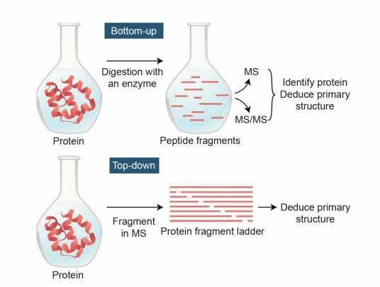
Why is it necessary to enzymatically digest proteins into peptides before mass spectrometry sequencing?expand_more
The larger the protein fragments, the lower the accuracy of mass spectrometry detection. Therefore, before mass spectrometry detection, proteins need to be digested into smaller peptides to improve detection accuracy. Generally, peptides with 6-20 amino acids are most suitable for mass spectrometry detection.
How to improve the efficiency of determining the sequence of modified peptide segments in protein sequencing?expand_more
Under current conditions, it is difficult for mass spectrometry to provide 100% sequence coverage of peptides. Some peptides may be lost during the process, and protein phosphorylation can inhibit trypsin digestion. Moreover, phosphorylated peptides are much less abundant than non-phosphorylated peptides, which may inhibit the mass spectrometry response. Therefore, efforts should be made to minimize non-phosphorylated peptides. Methods such as fractionation, IMAC, and antibody binding can be used. MALDI-TOF-MS can be used to determine the molecular weight of peptide segments; if the measured mass is 80 Da or its multiples higher than expected, phosphorylation can be inferred.
What could cause two amino acid residues to undergo Edman degradation simultaneously in the determination of protein N-terminal amino acid sequences?expand_more
When two amino acid residues are detected simultaneously in Edman degradation, it is possible that the protein is impure and contaminated with other proteins. If one of the amino acids is glycine, residual glycine from the buffer in the protein band may not have been completely removed, leading to this result.

For research use only, not intended for any clinical use.

Online Inquiry
Online Inquiry