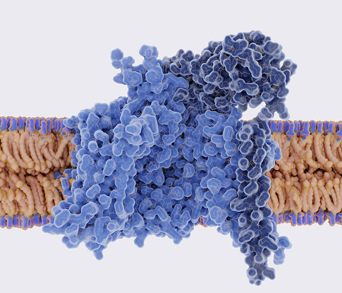
Pathogens or cancer cells that infect the human body release specific antigens, stimulating CD8+ naïve T cells to differentiate into effector cells. After the antigens in the body are cleared, effector T cells undergo rapid cell death, which is crucial for maintaining immune homeostasis. If effector T cells are not eliminated after the foreign pathogens are eradicated, it can lead to autoimmune responses that harm health. However, the mechanisms behind the death of effector T cells have remained poorly understood.
On September 11, 2024, Nature Cell Biology (IF 17.3) published a research paper titled "Ammonia-induced lysosomal and mitochondrial damage causes cell death of effector CD8+ T cells", which discovered that ammonia gradually accumulates inside CD8+ T cells during activation, ultimately leading to T cell death. This study reveals a novel mechanism for the rapid death of effector T cells.
For the first time, the authors introduced a new form of cell death called "ammonia-induced death." Ammonia-mediated cell death is a distinct form of cell death whose morphology differs from apoptosis, ferroptosis, and pyroptosis. This may be due to the different mechanisms that induce cell death. For example, apoptosis is triggered by intrinsic and extrinsic signal molecules that activate caspases 3 and 7; ferroptosis is initiated by ROS-induced excessive lipid oxidation in an iron-dependent manner; pyroptosis is triggered by the activation of gasdermins, which form pores in the plasma membrane from the inside out. However, ammonia-induced cell death is caused by ammonia, which damages lysosomes and mitochondria by consuming protons.
Select Service
Main Research Content
The research team's previous studies found that CD8+ T cells can clear harmful ammonia through the activation of the urea and citrulline cycles, aiding memory cell differentiation and long-term survival. During the activation and expansion of T cells, a large amount of ammonia is produced. If this ammonia is not fully cleared, it may accumulate in the body, eventually leading to T cell death. The authors hypothesize that accumulated ammonia during differentiation and proliferation could be a key factor in effector T cell death.
The authors established an animal model by transferring CD8+ T cells into C57BL/6J mice and then injecting Listeria expressing ovalbumin to activate the cells. They analyzed the changes in ammonia levels and related phenotypes in effector CD8+ T cells under pathogen infection conditions. In a mouse tumor model, the authors confirmed the occurrence of ammonia accumulation and lysosomal pH elevation, signs of ammonia-induced cell death, in tumor-infiltrating T cells.
 Figure 1: Ammonia-Induced Death of Effector CD8+ T Cells (Source: Reference 1)
The authors observed that during the early stages of infection, the ammonia levels within T cells showed a declining trend, which then gradually increased. By day 12 of infection, ammonia concentration within T cells reached its peak, and at this stage, the mortality rate of antigen-specific T cells significantly increased. This phenomenon suggests that the accumulation of ammonia may be closely related to the death of effector T cells. At this point, ammonia was not detected in the culture medium, and it was not until day 16 that ammonia was found in the supernatant of the culture medium, indicating that ammonia was produced by the T cells themselves.
Cell death was characterized by shrinkage and the appearance of numerous vacuoles. Further identification by the authors revealed that this type of death was distinct from caspase-dependent apoptosis, ferroptosis, and autophagy. Mitochondrial respiration and energy production depend on the proton gradient of the inner membrane. Therefore, the authors examined the impact of ammonia on the mitochondria in long-term activated effector CD8+ T cells.
 Figure 2: Ammonia Derived from Glutamine Causes Death of Effector T Cells (Source: Reference 1)
In long-term activated effector CD8+ T cells, ammonia levels were significantly increased, while the mitochondrial membrane potential was notably reduced. This indicates that mitochondrial energy production was inhibited. Furthermore, the copy number of mitochondrial DNA was significantly decreased, further reflecting the decline in mitochondrial function. The structure of the mitochondrial membranes and cristae was severely damaged, which may have profound effects on the metabolism and function of T cells. Subsequently, tumor mouse models were treated with the GLS1 inhibitor JHU083 (which inhibits ammonia production) or C381 (which lowers lysosomal pH). Both treatments were observed to suppress tumor growth and effectively prolong the survival time of the mice.
 Figure 3: Ammonia Storage in Effector CD8+ T Cells and Lysosomal Damage (Source: Reference 1)
These findings suggest that the accumulation of ammonia and its metabolic products are key mechanisms leading to the dysfunction of effector T cells. During activation, CD8+ effector T cells rely on glutamine metabolism to provide precursors for cell growth and proliferation. In the early stages of effector T cell proliferation, these cells use ammonia generated from glutamine metabolism to quickly enter anabolic pathways, preventing the buildup of ammonia within the cell.
However, in the later stages of effector T cell activation, cell proliferation gradually halts. Unlike memory T cells, effector T cells exhibit low expression of carbamoyl-phosphate synthetase 1 (CPS1), a key rate-limiting enzyme in the urea cycle. As a result, they are unable to convert ammonia into urea for extracellular excretion via the urea cycle, leading to the accumulation of ammonia inside the cell. Overexpression of CPS1 or the use of the ammonia scavenger 4-phenylbutyric acid significantly reduces effector T cell death.
 Figure 4: Ammonia Retention Leads to Mitochondrial Damage (Source: Reference 1)
Based on the unique death mechanism of effector T cells, the research team named this process "Ammonium-induced death" (AID). This also provides a new potential anti-cancer mechanism, wherein enhancing anti-tumor immunity by inhibiting ammonia-induced death of effector T cells could be a strategy. The authors further transplanted effector T cells pretreated with JHU083 or C381 into mice, and the results showed that these treatments also enhanced T cell persistence, effectively controlled tumor growth, and improved the survival rate of the mice.
 Figure 5: Damaged Mitochondria Cannot Be Cleared Through Autophagy (Source: Reference 1)
By combining inhibitors that suppress ammonia-induced death with existing immunotherapy drugs, or by pre-treating T cells with ammonia death inhibitors, new treatment options may be provided for cancer patients.
 Figure 6: Ammonia Death Blockade Enhances Cancer T Cell Therapy (Source: Reference 1)
In summary, the characteristics of ammonia-induced death include:
- It is dependent on the accumulation of ammonia ions rather than oxidative stress or iron metabolism disorders;
- It primarily occurs in effector CD8+ T cells with high metabolic activity, rather than in other cell types;
- Unlike apoptosis and necrotic cell death, it is a regulated form of new cell death.
This discovery highlights the complexity of immune regulation in the human body, where activation and inhibition coexist in the same process: in rapidly proliferating T cells, mitochondria break down glutamine to promote cell proliferation while releasing ammonia. As ammonia gradually accumulates, it enters the lysosome, increasing the lysosomal pH and impairing its function. Since the cell cannot effectively process ammonia, these substances accumulate inside the cell, particularly within mitochondria, ultimately causing severe mitochondrial damage, leading to effector T cell death.
Inhibiting glutamine breakdown or preventing lysosomal alkalinization can effectively prevent ammonia-induced T cell death. This strategy not only enhances the understanding of ammonia-induced death but also improves the effectiveness of T cell-based anti-tumor immunotherapies, providing new ideas and possibilities for future cancer treatments.
Lysosomal pH elevation can serve as a marker for ammonia death. Although ammonia death has been demonstrated using effector T cells as a model, these findings may not be common, as ammonia dilution via lysosomal division and biosynthesis can prevent ammonia death in normal cells. In this regard, ammonia-induced cellular damage may be more related to chronic conditions, whereas apoptosis, ferroptosis, and pyroptosis may be more associated with acute conditions.
The formation of ammonia death depends not only on the buffering role of lysosomes in ammonia but also on the cell's strong anti-ammonia capacity. Even though the urea cycle (the primary mechanism for ammonia detoxification) is not common in tissue cells, extrahepatic cells can fix ammonia into the form of hypoxanthine urate through the purine synthesis pathway or use ammonia to produce polyamines (putrescine, spermidine, and spermine). Therefore, cells may not undergo ammonia death after short-term stimulation, which may explain why ammonia death is not easily observed. However, chronic ammonia accumulation can lead to cellular dysfunction, causing cell damage or even ammonia death. Thus, blocking ammonia production may be a useful strategy for disease intervention.
In this study, the authors found that inhibiting glutamine breakdown during certain early stages of T effector cells enhances anti-tumor immunity. This may be because reduced glutamine breakdown hampers the T effector cell's tricarboxylic acid cycle during rapid proliferation, thereby decreasing ATP and ROS production, which promotes the early differentiation of T cells into memory CD8+ T cells. Additionally, brief GLS1 inhibition or lysosomal acidification may induce chromatin accessibility changes and retain epigenetic characteristics, which may help CD8+ T cells persist in tumors.
Overall, this study reveals a new form of cell death—ammonia death—where blocking ammonia death enhances T cell persistence. This discovery not only broadens the understanding of types of cell death but also provides new ideas and strategies for cancer therapy.
Comparison with Ferroptosis and Cuproptosis
In recent years, cell death-related research hotspots, such as ferroptosis and cuproptosis, have gained significant attention in national research funding applications. However, their mechanisms of occurrence differ.
Characteristics of Ferroptosis:
- It depends on the accumulation of iron ions and lipid peroxidation reactions, leading to membrane rupture;
- It is widely present in different types of cells;
- It is induced by inhibiting glutathione synthesis and the antioxidant activity of glutathione-dependent peroxidase (GPX4);
- Lipophilic free radical scavengers can prevent lipid peroxidation accumulation and specifically inhibit ferroptosis;
- There is no single universal ferroptosis pathway; many different metabolites and proteins can initiate, promote, and regulate ferroptosis, but none are strictly essential;
- Cellular sensitivity to ferroptosis can be altered by various signals and transcriptional networks.
Characteristics of Cuproptosis:
- It depends on the accumulation of copper ions, leading to the aggregation of acylated proteins and a reduction in iron-sulfur cluster proteins;
- It mainly occurs in cells that rely on mitochondrial respiration, where the lipoacylation modification of key mitochondrial metabolic enzymes is closely linked to copper-induced cell death;
- It is induced by disrupting the ubiquitin-proteasome system and the tricarboxylic acid cycle;
- Copper ion uptake genes are associated with copper death tolerance;
- Copper death is related to the occurrence and progression of various diseases, including breast cancer, lung cancer, neurodegenerative diseases, Wilson's disease, obesity, and cardiovascular diseases.
Ammonia death, ferroptosis, and cuproptosis are all newly discovered forms of cell death that depend on the accumulation of specific ions and induce cell death through different molecular pathways. Understanding the differences in these cell death mechanisms can help develop new treatment strategies to enhance cellular survival, especially in applications such as cancer immunotherapy.
References
- Zhang H, et al. Ammonia-induced lysosomal and mitochondrial damage causes cell death of effector CD8+ T cells. Nat Cell Biol. Published online September 11, 2024. doi:10.1038/s41556-024-01503-x
- Tang K, et al. Ammonia detoxification promotes CD8+ T cell memory development by urea and citrulline cycles. Nat Immunol. 2023;24(1):162-173. doi:10.1038/s41590-022-01365-1