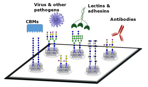
Biological microarrays have emerged as crucial tools for efficiently and comprehensively extracting pertinent information. Concurrently, as glycobiology and glycomics research advances, a novel type of biochip—glycan microarrays—is progressively being established as a pioneering approach in the realm of glycobiology and glycomics.
Glycans, encompassing monosaccharides, oligosaccharides, or polysaccharides linked to proteins or lipids, permeate the biological landscape, imparting profound significance to diverse physiological processes. Notably, cell surface glycans play a dual role: they facilitate normal cellular recognition, adhesion, and intercellular communication, while also bearing pivotal relevance in uncovering cellular malfunctions and pathogen interactions. The aberrant glycan structures and activities seen in processes like cellular carcinogenesis and the recognition of host cells by viruses and bacteria accentuate the importance of studying alterations in cell surface glycans. However, the intricate nature of polysaccharides—marked by variations in monosaccharide composition, stereochemical configurations, glycosidic bond types, and complex biosynthetic pathways—poses formidable challenges to glycan investigation.
Principle of Glycan Microarrays
The concept of glycan microarrays, built upon the foundation of specific interactions between glycans and glycan-binding proteins (GBPs), has captured significant attention in scientific exploration. Glycan microarrays entail the immobilization of diverse glycans, each boasting unique structures, onto chemically-modified substrates via covalent or non-covalent bonds. Consequently, these arrays serve as platforms to evaluate and dissect samples, encompassing both glycan-binding proteins and glycan probes themselves. Molecules exhibiting precise interactions with the immobilized glycans become anchored, while non-specific entities are eliminated through rinsing procedures. Employing techniques such as fluorescence staining permits facile and expeditious identification of molecules exhibiting discernible interactions, ultimately playing a pivotal role in unraveling the structural and functional attributes of glycan-binding proteins.
Utilizations of Glycan Microarrays
Glycan microarrays have emerged as a versatile methodology for scrutinizing alterations in glycosylation within intricate biological specimens. Their utility extends to elucidating the interplay between glycans and glycan-binding proteins (GBPs), as well as facilitating kinetic assessments of glycan-protein interactions. The scope of glycan microarray applications has broadened, encompassing the discrimination of interactions with viruses, bacteria, cells, and dynamic responses within living cells. Furthermore, these arrays harbor the potential to uncover utilitarian glycans, characterize enzymatic processes, identify pathogens, and oversee an array of molecular interactions involving glycans.
 Figure 1. Schematic diagram of the various applications of glycan microarrays
Glycan Microarray Analysis Services
We provide glycan microarray analysis services to aid researchers in deciphering protein-glycan interactions. The glycan microarray platform enables the analysis of various biological samples, such as proteins, antibodies, cells, cell lysates, serum, vesicles, bacteria, or viral particles. Our experts can assist in customizing a plan tailored to your research needs. From study design to product and service selection, sample collection, reporting, and biological interpretation, our experts guide you at every step. Your research success is our goal.
Determining Glycan-Binding Protein (GBP) Specificity
Carbohydrates manifest in various forms, including free oligosaccharides, glycolipids, glycoproteins, and other glycoconjugates, within nearly all life forms. A subset of carbohydrates, referred to as glycans, participates in diverse biological events, such as disease mechanisms, humoral immunity, and HIV glycan shielding. Glycans also serve as therapeutic targets for cancer immunotherapy. The complex nature of glycans mediates intricate cell signaling, often involving multivalent interactions. For a given GBP, multiple binding sites may exist, each with its own specificity and binding affinity. Glycan-binding proteins can be categorized as extracellular or intracellular. External GBPs (e.g., plant lectins) recognize mammalian glycans from various organisms, while internal GBPs (e.g., Siglecs) recognize mammalian glycans from the same organism. Glycan microarray technology evaluates binding interactions between glycans and GBPs, offering a comprehensive landscape of glycan-binding specificity.
To ascertain GBP binding specificity, we employ existing or client-customized glycan microarrays. During this process, we first immobilize glycans onto a solid-phase surface using ultra-low autofluorescence high-quality glass slides. Single glycans from natural sources or chemically/enzymatically synthesized ones are then covalently linked to the substrate, creating microarray chips for downstream analysis. During binding assays, a proprietary blocking buffer is used to seal the microarray, followed by the introduction of GBPs. Detection reagents, such as fluorescently labeled antibodies, are employed to capture GBP binding. At assay conclusion, fluorescence signals are quantified in relative fluorescence units (RFUs). Various bioinformatics tools are utilized to analyze data and determine GBP binding specificity.
Our Glycan Microarrays Analysis Process

Advantages of Glycan Microarrays
High Throughput: Glycan microarray analysis requires minimal sample volume yet supports high throughput. Thousands of distinct samples can be analyzed in parallel on a small chip.
Broad Applicability: Glycan microarrays, generated by covalently or non-covalently immobilizing synthetically derived or natural glycans on substrates like glass, polyvinylidene fluoride, or nitrocellulose, find applications in identifying sugar-recognizing proteins, characterizing glycoprotein binding specificity, pathogen detection, drug target screening, and more.
Comprehensive: They facilitate the study of interactions with a diverse range of glycans in a single assay.
How Does A Glycan Microarray Work
The functioning of a glycan microarray hinges on the immobilization of an array of diverse glycans, or carbohydrate molecules, onto a solid substrate, which can range from a glass slide to a microchip. These immobilized glycans serve as molecular probes to scrutinize their interactions with a spectrum of target molecules, encompassing proteins, antibodies, cells, viruses, and other biomolecules. The primary objective is to comprehend and delineate the binding preferences and specificities of these target molecules towards different glycans.
Outlined below is the typical procedure through which a glycan microarray operates:
Glycan Immobilization: An assortment of meticulously characterized glycans is affixed to predefined locations on the microarray substrate. Each distinct spot on the array corresponds to a unique glycan structure. These glycans may be derived from natural sources or synthesized to replicate particular structures.
Target Interaction: The microarray is exposed to a solution containing the target molecules under investigation. These targets encompass a gamut of entities, including proteins, antibodies, lectins (proteins that exhibit specific glycan affinities), and other molecules bearing glycan-binding propensities.
Binding Assay: The target molecules initiate interactions with the immobilized glycans adorning the microarray surface. Should a target molecule bind to a specific glycan structure, a complex ensues that remains tethered to the microarray spot.
Detection: Multiple detection methodologies can be deployed to visualize these binding interactions. A common strategy involves labeling the target molecules with fluorescent or radioactive markers, thereby facilitating the detection of their binding to the glycan-occupied spots. The extent of binding can subsequently be gauged by quantifying the intensity of fluorescence or radioactivity.
Data Analysis: The resultant data, often presented as either images or numerical values, unveil the glycans with which the target molecules interact. The vigor and specificity of these interactions can be ascertained by scrutinizing fluorescence intensity or analogous detection signals.


