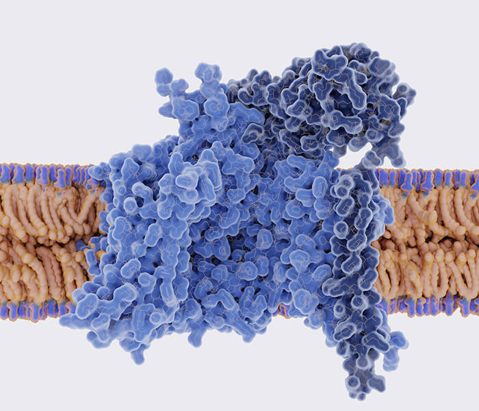
The central metabolic pathway is a core set of metabolic reactions in organisms responsible for converting external carbon sources into energy, cellular structures, and precursors for biomolecules. Central metabolism not only supports the basic life activities of an organism, but also provides cells with the metabolic substances necessary to sustain growth, reproduction, and response to environmental changes. These metabolic pathways, including glycolysis, the tricarboxylic acid cycle (TCA cycle), the pentose phosphate pathway, as well as fatty acid synthesis and amino acid synthesis, constitute a complex and highly regulated network. In-depth analysis of the operation mechanisms of these pathways is important for understanding the basic life processes of organisms, developing new biotechnological tools, and treating metabolism-related diseases.
In this article, we will explore how to analyze central metabolic pathways from multiple perspectives, including experimental methods, computational modeling, and systems biology approaches, and demonstrate the application of these methods through example analyses. Through the in-depth study of these methods, we can have a more comprehensive understanding of the energy flow, substance conversion, and metabolite regulation mechanisms in metabolic processes.
 Upper central carbon metabolism (Long CP et al., 2016)
Determination of enzyme activity
Spectrophotometry (taking hexokinase as an example)
The rate of enzyme catalysis is calculated using Beer-Lambert's law by detecting the change in absorbance of a product or substrate in an enzymatic reaction at a specific wavelength (e.g., NADPH absorbance at 340 nm).
- Preparation of reaction system: reaction buffer (pH 7.4, containing Mg, ATP and NADP⁺).
- Substrate: glucose (final concentration 5 mM).
- Auxiliary enzyme: glucose -6- phosphate dehydrogenase (G6PDH, used to convert the product G6P into 6- phosphate gluconolactone and generate NADPH at the same time).
- Start the reaction: add hexokinase extract, mix it immediately and put it in a spectrophotometer. Monitor the change of NADPH absorbance at 340 nm with time (recorded every 30 seconds for 5 minutes).
- Data analysis: According to the change rate of absorbance (δ A/min) and the molar extinction coefficient of NADPH (ε = 6.22 mmcm), the enzyme activity (unit: μmol/min/mg protein) was calculated.
- Key instruments: ultraviolet-visible spectrophotometer, constant temperature cuvette.
Radiolabeling method (taking citrate synthase as an example)
Using isotope-labeled substrates (e.g., 14C), the accumulation of radioactive signals in the product is tracked, and the enzyme activity is quantified by the intensity of the radioactivity.
- Isotopic labeling substrate: C-labeled oxaloacetic acid (OAA) was mixed with A(Acetyl-CoA.
- Reaction termination and separation: After a certain time, trichloroacetic acid (TCA) is added to terminate the reaction. The product citric acid was separated by thin layer chromatography (TLC) or HPLC.
- Radioactivity detection: collect citric acid components and measure the radioactivity of C with liquid scintillation counter.
- Calculation of enzyme activity: radioactivity count/(reaction time × protein concentration).
- Limitations: radioactive waste needs to be treated, and the operation safety is required to be high.
Metabolite concentration analysis
Separation of metabolites using reversed-phase columns, characterization based on retention time, and quantification based on peak area (e.g., ATP/ADP).
- Sample quenching: the cell culture was quickly poured into methanol/water (80:20) precooled by liquid nitrogen and stored at -80℃.
- Metabolite extraction: the cells were broken by ultrasound and centrifuged (15,000×g, 10 min) to get the supernatant. Vacuum drying and redissolving in HPLC mobile phase (such as phosphate buffer).
- Chromatographic conditions: chromatographic column: C18 reversed-phase column (4.6×250 mm). Mobile phase: gradient elution (0.1 M KH₂PO₄, pH 6.0 → methanol). Detector: ultraviolet detector (254 nm, ATP/ADP characteristic absorption).
- Quantitative analysis: the concentration is calculated by the retention time and peak area of the standard.
GC-MS analysis of pyruvate
Volatile metabolites were separated by gas chromatography and identified and quantified by mass spectrometry after ionization based on mass-to-charge ratio (m/z) and fragmentation ion spectra (e.g., pyruvic acid needs to be detected after derivatization).
- Derivatization treatment: the sample reacts with methoxyamine hydrochloride (MOX) to block ketone groups; Then the hydroxyl group was derivatized with BSTFA (silanization reagent).
- Mass spectrometry parameters: ion source: EI(70 eV), full scanning mode (m/z 50-600). Quantitative ions: characteristic fragments of pyruvate derivatives (such as m/z 147).
Isotope tracing
Stable isotope (e.g., ¹³C)-labeled substrates (e.g., glucose) are introduced into the metabolic system, and the distribution of the isotopes in the metabolites is tracked by mass spectrometry, which is combined with mathematical models to calculate metabolic fluxes.
- Labeling strategy: use U-C glucose (all carbon atoms are C).
- Cell treatment: the cancer cells were cultured in a culture medium containing C glucose for 4 hours. Quickly quench and extract metabolites (lactic acid, TCA cycle intermediates).
- Mass spectrometry analysis: lactic acid (m/z 89→90, C label increased by 1 Da) and TCA metabolites (such as the distribution of C isotope isomers of citric acid) were detected by LC-MS.
- Flux calculation: use software (such as Isotopo) to analyze the flow direction of carbon atoms and quantify the ratio of glycolysis (lactic acid production) to mitochondrial oxidation (CO release).
Multi-group integration technology
Un-targeted metabonomics (LC-MS/MS)
Simultaneous analysis of hundreds of metabolites based on liquid chromatography separation and high sensitivity detection by mass spectrometry, combined with database annotation.
- Sample preparation: plasma/cell lysate was precipitated with acetonitrile, and the supernatant was dried and then dissolved in methanol.
- Chromatographic conditions: reversed-phase column (such as Zorbax Eclipse Plus C18), mobile phase of 0.1% formic acid water (a) and acetonitrile (b), gradient elution (5% B → 95% B, 20 min).
- Mass spectrometry parameters: high-resolution mass spectrometry (such as Q-Exactive), positive/negative ion mode switching, data-dependent acquisition (DDA).
- Data analysis: using XCMS or MetaboAnalyst for peak alignment and compound annotation (based on m/z and MS/MS libraries).
Analysis of phosphorylated protein group
Enrichment of phosphorylated peptides (e.g., TiO2 microcolumns) and identification of phosphorylation sites by mass spectrometry reveal the mechanism of enzyme activity regulation.
- Enrichment of phosphorylated peptides: using TiO2 microcolumn to selectively bind phosphorylated peptides.
- Mass spectrometry analysis: after liquid phase gradient separation, Orbitrap mass spectrometry collected MS/MS spectrum.
- Data analysis: Software (such as MaxQuant) matched the database of phosphorylation sites to quantify the changes of phosphorylation level.
Single cell metabonomics (CyTOF)
Single-cell surface proteins were labeled with metal-tagged antibodies, and metal signals were detected by inductively coupled plasma mass spectrometry to resolve the heterogeneity of metabolism-related protein expression.
- Metal-labeled antibody labeling: incubating single cell suspension with lanthanide-labeled antibodies (such as anti -GLUT1 and anti -HK2).
- Mass spectrometry flow detection: Cells pass through inductively coupled plasma (ICP) one by one, and metal labels are ionized and detected.
- Data analysis: t-SNE and clustering were used to identify metabolic heterogeneity subgroups.
Living cell imaging
Gene-encoded fluorescent protein sensors undergo conformational changes upon binding to metabolites (e.g., NAD⁺/NADH), reflecting dynamic metabolite concentrations through fluorescence intensity ratios.
- Sensor transfection: iNap plasmid was transfected into cells by electroporation or lentivirus.
- Dual-channel imaging: excitation wavelength: 405 nm(NADH bound state) and 488 nm (total sensor). Emission wavelength: 500-550 nm.
- Ratio calculation: NAD⁺/NADH ratio =(488 nm signal-405 nm signal)/405 nm signal.
MFA
Integration of isotope labeling data with metabolic network modeling to quantify metabolic flux distributions (e.g., the ratio of glycolysis to PPP contribution) through mass conservation and isotope balance equations.
- Labeled Substrate Treatment: Cells or tissues are cultured in medium containing ¹³C-labeled substrate (e.g., [U-¹³C]-glucose). Control temperature, CO₂ concentration and labeling time.
- Metabolic quenching and sample collection: Rapid quenching: add pre-cooled methanol/water mixture and liquid nitrogen flash freeze. Metabolite extraction: metabolite extraction using acetonitrile/methanol mixture, centrifugation to remove proteins.
- Isotope Isomer Detection: LC-MS: use a HILIC or C18 column with gradient elution. Detection of isotopic isomers (e.g. [M+0], [M+1]).
- Metabolic network modeling: Construct stoichiometric matrices to describe substrate-product relationships. Define isotope transfer pathways and construct isotope mapping matrices.
- Flux Calculation and Optimization: Minimize differences between experimental data and model predictions. Calculate metabolic flux values using software tools.
- Data Visualization & Interpretation: Heatmaps showing flux changes. Metabolic pathway maps overlay flux values.
Services You May Be Interested In:
Additional Resources:
Examples of applications
Development of biomarkers
13C-MFA not only reveals the metabolic profile of tumor cells, but also helps to identify metabolites associated with tumor progression. For example, certain metabolic intermediates such as lactate, pyruvate, and glutamate may become potential tumor markers. By 13C-labeled metabolic tracking, the production rate of these metabolites can be monitored, revealing the level of metabolic activity of the tumor.
- BRAF mutation (especially BRAF V600E mutation) is considered as one of the key drivers of melanoma, which affects cell proliferation, differentiation and apoptosis by activating MAPK signaling pathway. The combination therapy of BRAF inhibitor (such as Verofibril) and MEK inhibitor shows certain curative effect, but the development of drug resistance limits the long-term therapeutic effect. HP 13C- pyruvate and 13C-MRS fluxes were used to evaluate the metabolic response of BRAF/MEK inhibitors to melanoma xenografts. In YUMM1.7 xenograft, the combination of BRAF and MEK inhibitor delayed the growth of tumor more significantly than single drug. Through the evaluation of p-ERK level after 4 hours, the response of all treatment groups showed a decrease in p-ERK level. However, only the decrease of p-ERK in MEK inhibition group was statistically significant, indicating that p-ERK as a biomarker could not fully predict the treatment response. The metabolic study of HP 13C- pyruvate showed that the ratio of lactic acid to pyruvate did not change significantly 24 hours after BRAF/MEK inhibitor treatment. However, in A375 melanoma xenograft model, the ratio of HP pyruvate to lactic acid increased significantly after BRAF inhibition treatment. This difference may be related to the change of tumor microenvironment or the influence of immune cells. In MEKi treatment group, the expression of MCT1 increased unexpectedly, but no significant effect of BRAF or MEK inhibition on the exchange of pyruvate and lactate was observed. After 13C- glucose labeling, the production of lactic acid decreased significantly, which indicated that the glycolytic activity of tumor cells might decrease. GLUT-1 expression did not change after BRAF/MEK inhibition, suggesting that the decrease of glycolysis may be related to glycolytic enzyme activities (such as hexokinase and pyruvate kinase dehydrogenase). In contrast, the metabolism of 13C- glutamine did not show significant changes, indicating that BRAF/MEK inhibition had little effect on glutamine pathway, especially in this model, glutamine pathway was independent. This study shows that HP 13C- pyruvate and 13C- glucose flux omics can be used as effective markers to identify the inhibitory response of BRAF/MEK (Farah C et al., 2022).
Research and optimization of bioenergy
Metabolic analysis is critical to the development of biofuels and other renewable energy sources. By analyzing how microorganisms metabolize using different carbon sources, researchers can optimize microbial metabolic pathways to improve the efficiency of biofuel (e.g., ethanol, butanol, etc.) production and advance the development of green energy.
- Glycerol is a by-product of biodiesel, and converting it into valuable chemicals (such as acetyl alcohol) is helpful to improve the economy of biofuels. Scientists analyzed the metabolic bottlenecks of different strains (including production strain HJ06 and non-production strain HJ06C) in the production of acetol by 13C-MFA, and found that NADPH regeneration was the key to improve the production of acetol. In this study, HJ06 strain successfully produced 0.91 g/L acetol from glycerol, while the control strain HJ06C did not produce acetol. Through 13C tracer experiment, it was found that the flux of acetate was higher than that of TCA, and the relative flux of pentose phosphate (PP) pathway was lower. The supply of NADPH in H06 strain is insufficient, which becomes the bottleneck of acetol production. Through overexpression of nadK gene, the supply of NADPH was further improved. The results showed that the yield of acetyl alcohol of HJ06N strain was 65% higher than that of HJ06, which indicated that the regeneration of NADPH was an effective optimization direction for the overproduction of acetyl alcohol. Through 13C-MFA, it was determined that the metabolic flux of strain HJ06N shifted slightly between glycolytic pathway and acetol biosynthesis pathway, showing a slightly Qualcomm amount from glycolytic pathway to acetol production pathway. The fluxes of HJ06 and HJ06N strains through pentose phosphate pathway (PP pathway) and tricarboxylic acid cycle (TCA cycle) are very similar. In HJ06N, the trans-hydrogen flux from NADH to NADPH increased by about 1.4 times, indicating that the regeneration of NADPH was enhanced. Comparing the expression profiles of different strains, it was found that zwf and gnd were down-regulated and sucA and gltA were up-regulated, which was consistent with the flux change. The comparison of gene expression profiles among HJ06N/HJ06, HJ06P/HJ06 and HJ06PN/HJ06 shows that there is no significant difference in the regulation of other genes, indicating that most metabolic changes are mainly concentrated in the pathway related to NADPH production and acetohydrin synthesis (Yao R et al., 2019).
Basic biological research
Metabolism is the basis of life activities within cells. Metabolic analysis can provide insights into how cells utilize nutrients to sustain life, produce energy, and synthesize cellular components. Through meticulous research, the Metabolic Analysis Center can reveal how different organisms regulate metabolic networks in different environments, thus helping to understand the fundamentals of life.
- P. aeruginosa activates different metabolic pathways under different carbon sources (e.g. acetate and glycerol). Specifically, under acetate conditions, the expression of three key enzymes (isocitrate dehydrogenase, isocitrate lyase, etc.) is increased, and these enzymes compete with each other for the substrate isocitrate. Growth of P. aeruginosa on acetate showed changes in the composition of the electron transport chain, especially in the terminal oxidase. The expression of genes encoding terminal oxidases was significantly increased under acetate conditions, whereas the expression of another class of oxidase genes (cox) was increased under glycerol conditions.These alterations in ETC suggest that the cells optimize energy production by using different oxidases. Under acetate growth conditions, it was found that the denitrification mechanism of P. aeruginosa underwent a significant aerobic induction. Denitrification is usually associated with hypoxic conditions, but it was shown that the denitrification system may also be initiated under aerobic conditions, possibly as a mechanism to restore redox homeostasis. P. aeruginosa grew at a faster rate under acetate conditions than under glycerol conditions. In order to maintain redox homeostasis, the cells excrete some oxidized metabolic intermediates through "spillover metabolism". Spillover metabolism may be a metabolic strategy by which cells can maximize their growth rate. The study suggests that the P. aeruginosa transcriptional regulator Anr may be active even in the presence of oxygen, regulating the expression of denitrification-related genes. This finding hints at the adaptability of Anr in the bacterial response to changes in oxygen. This study reveals how P. aeruginosa maintains growth and redox homeostasis by adjusting metabolic pathways and denitrification mechanisms under different growth conditions, especially on infection-related carbon sources (Dolan SK et al., 2020).
 Biochemical pathways involved in central carbon catabolism in P. aeruginosa PAO1 (Dolan SK et al., 2020).
Environmental and ecological studies
Soil is one of the largest carbon reservoirs on Earth, storing large amounts of organic carbon. Microorganisms convert this organic carbon into carbon dioxide for release into the atmosphere by decomposing, for example, plant residues and animal organic matter, or fix the carbon by synthesizing new organic matter. Understanding the metabolic processes of carbon sources in soil can help to reveal the source, transformation and storage mechanisms of soil carbon, so as to better predict and manage the release and fixation of soil carbon, and thus mitigate or exacerbate climate change.
- Understand how soil microbial communities metabolically process different carbon sources (e.g., cellulose, glucose, xylose, amino acids from plants, etc.) and analyze the mechanisms of these transformations and the amount of time that carbon remains in the soil. Nine carbon sources were chosen to represent common classes of compounds found in soils. It was found that 69 of the 138 metabolites were significantly enriched in 13 C. Most of the enriched metabolites were involved in central metabolic pathways such as amino acid synthesis, catabolism, and DNA synthesis. Different carbon sources led to different 13C enrichment patterns of the metabolites, especially the enrichment rates and metabolite profiles differed between soluble and insoluble carbon sources. Significant changes in metabolite profiles and microbial community composition occurred over time. Although the metabolite profiles of the different carbon sources did not differ significantly at first, the metabolite enrichment gradually decreased as the metabolites were consumed. The 13C enrichment of specific metabolites by certain carbon sources (e.g. aromatic compounds) revealed ecological differences in community metabolism. For example, the production of benzoic acid and salicylic acid reflected the effects of amino acid catabolism and glucose biosynthesis, respectively. The 13C enrichment of certain metabolites involved in osmoregulation (e.g., glycine betaine and ectoin) also showed significant differences between carbon sources. These differences may reflect cellular adaptive mechanisms in response to metabolic stresses such as high metabolic activity or aromatic oxidation. Disaccharides (e.g., maltose and alginate) also showed different 13C enrichment patterns in the metabolism of different carbon sources, suggesting that the metabolic pathways of different carbon sources may involve the storage and release of carbon and energy (Wilhelm RC et al., 2022).
References
- Farah C, Neveu MA, Yelek C, Bouzin C, Gallez B, Baurain JF, Mignion L, Jordan BF. "Combined HP 13C Pyruvate and 13C-Glucose Fluxomic as a Potential Marker of Response to Targeted Therapies in YUMM1.7 Melanoma Xenografts." Biomedicines. 2022;10(3):717. doi: 10.3390/biomedicines10030717
- Yao R, Li J, Feng L, Zhang X, Hu H. "13C metabolic flux analysis-guided metabolic engineering of Escherichia coli for improved acetol production from glycerol." Biotechnol Biofuels. 2019 ;12:29. doi: 10.1186/s13068-019-1372-4
- Dolan SK, Kohlstedt M, Trigg S, Vallejo Ramirez P, Kaminski CF, Wittmann C, Welch M. "Contextual Flexibility in Pseudomonas aeruginosa Central Carbon Metabolism during Growth in Single Carbon Sources." mBio. 2020 ;11(2):e02684-19. doi: 10.1128/mBio.02684-19
- Wilhelm RC, Barnett SE, Swenson TL, Youngblut ND, Koechli CN, Bowen BP, Northen TR, Buckley DH. "Tracing Carbon Metabolism with Stable Isotope Metabolomics Reveals the Legacy of Diverse Carbon Sources in Soil." Appl Environ Microbiol. 2022 ;88(22):e0083922. doi: 10.1128/aem.00839-22