What is Phosphatidic Acid?
Phosphatidic acid (PA) is a crucial lipid molecule found in biological systems. Structurally, it consists of a glycerol backbone with two fatty acid chains esterified at positions sn-1 and sn-2, and a phosphate group esterified at position sn-3. This unique structure makes PA an amphipathic molecule, meaning it has both hydrophobic (fatty acid chains) and hydrophilic (phosphate group) regions.
PA is a key intermediate in lipid metabolism and serves as a precursor for the biosynthesis of other phospholipids, such as phosphatidylcholine and phosphatidylethanolamine. It is synthesized via several pathways, including the Kennedy pathway, de novo synthesis, and the phospholipase D pathway.
Beyond its role in lipid metabolism, PA acts as a signaling molecule involved in various cellular processes. It regulates membrane dynamics, vesicle trafficking, and cytoskeletal organization. Moreover, PA participates in signal transduction pathways by activating protein kinases, phospholipases, and other signaling molecules. These signaling events contribute to cell proliferation, differentiation, and survival.
Overall, phosphatidic acid plays diverse roles in cellular physiology and is essential for maintaining cellular homeostasis and function. Its multifunctional nature makes it a subject of intense research interest in fields ranging from lipid biochemistry to cell biology and biomedical sciences.
Chemical Properties of Phosphatidic Acid
Molecular Structure
At its core, phosphatidic acid comprises a glycerol backbone esterified with two fatty acid chains at the sn-1 and sn-2 positions, and a phosphate group at the sn-3 position. This tripartite structure renders PA amphipathic, with hydrophobic tails derived from fatty acids and a hydrophilic head group constituted by the phosphate moiety. The amphipathic nature of PA facilitates its interaction with both hydrophilic and hydrophobic environments, crucial for its diverse biological functions.
Functional Groups and Chemical Properties
The presence of ester bonds between the glycerol backbone and fatty acid chains confers stability to phosphatidic acid molecules, while the phosphate group imparts a negative charge to the head group, influencing its chemical behavior. This charge property enables PA to participate in electrostatic interactions with other molecules, such as proteins and metal ions, modulating its functional roles in cellular processes.
Stereochemistry and Isomeric Forms
Phosphatidic acid exists in various isomeric forms, depending on the stereochemistry of fatty acid chains and the position of double bonds within these chains. Isomeric diversity contributes to the structural heterogeneity of PA species, influencing their physical properties and biological functions. Additionally, the configuration of fatty acids at the sn-1 and sn-2 positions confers distinct properties to individual PA molecules, impacting their interactions with membrane proteins and lipid bilayers.
Conformational Dynamics
The conformational flexibility of phosphatidic acid molecules plays a pivotal role in membrane dynamics and lipid-protein interactions. PA molecules can adopt different conformations in response to changes in environmental conditions, such as pH, temperature, and lipid composition. These conformational changes influence the biophysical properties of membranes, including curvature, fluidity, and permeability, thereby regulating cellular processes such as vesicular trafficking and membrane fusion events.
What is The Difference Between Phospholipid and Phosphatidic Acid?
Phospholipids constitute a diverse group of lipids characterized by a glycerol backbone esterified with two fatty acid chains at the sn-1 and sn-2 positions, and a phosphate group at the sn-3 position. The phosphate group is further linked to a polar head group, such as choline, ethanolamine, serine, or inositol, imparting distinctive properties to different phospholipid species. Phospholipids serve as integral components of cellular membranes, contributing to membrane structure, fluidity, and function.
Phosphatidic acid, on the other hand, represents a specific subclass of phospholipids distinguished by its singular phosphate head group and pivotal role in lipid metabolism pathways. Structurally, PA comprises a glycerol backbone esterified with two fatty acid chains at the sn-1 and sn-2 positions, and a phosphate group at the sn-3 position. Unlike other phospholipids, PA lacks a polar head group, rendering it amphipathic with a net negative charge at physiological pH.
Key Differences:
- Presence of Polar Head Group: Phospholipids possess a polar head group, which imparts unique chemical properties and biological functions to different phospholipid species. In contrast, phosphatidic acid lacks a polar head group, contributing to its amphipathic nature and distinctive role in cellular physiology.
- Biological Functions: Phospholipids serve diverse functions in cellular membranes, including structural support, cell signaling, and membrane trafficking. Phosphatidic acid, while also contributing to membrane structure, plays a central role in lipid metabolism regulation, signaling transduction, and cellular responses to environmental cues.
- Metabolic Significance: Phospholipids are ubiquitous constituents of cellular membranes, essential for maintaining membrane integrity and function. Phosphatidic acid serves as a precursor for the biosynthesis of other phospholipids and bioactive lipid molecules, regulating lipid metabolism pathways and cellular homeostasis.
Physiological and Biological Functions of Phosphatidic Acid
Phosphatidic acid (PA) serves as a pivotal regulator of diverse physiological processes and biological functions within biological systems. Its multifaceted roles encompass membrane composition, signal transduction, lipid metabolism regulation, and modulation of cellular functions.
Cellular Membrane Composition and Signaling
Phosphatidic acid exerts profound effects on cellular membrane composition and dynamics, exerting influence over membrane curvature, lipid bilayer fluidity, and protein-lipid interactions. As a key constituent of lipid bilayers, PA contributes significantly to membrane structure and organization, facilitating membrane remodeling processes such as endocytosis, exocytosis, and vesicular trafficking. Moreover, PA acts as a signaling molecule, transducing extracellular signals into intracellular responses through activation of protein kinases, phospholipases, and second messenger cascades. By modulating the function and localization of membrane-associated proteins, PA regulates diverse cellular processes including cell proliferation, differentiation, and survival.
Influence on Membrane Dynamics
Phosphatidic acid's amphipathic nature allows it to insert into lipid bilayers, altering membrane curvature and influencing membrane morphology. By promoting membrane bending and fusion events, PA facilitates the formation of membrane structures such as endocytic vesicles, Golgi cisternae, and autophagosomes. Additionally, PA regulates lipid raft formation, membrane domain organization, and protein sorting within cellular membranes, contributing to membrane compartmentalization and function.
Role in Intracellular Signaling
Phosphatidic acid serves as a versatile signaling molecule involved in the transduction of extracellular signals into intracellular responses. PA-mediated signaling pathways encompass a diverse array of cellular processes, including growth factor signaling, stress responses, and cytoskeletal rearrangements. Key mechanisms underlying PA-mediated signaling include activation of protein kinases, phospholipases, and second messenger cascades, leading to the modulation of gene expression, cytoskeletal dynamics, and cell motility. Moreover, PA interacts with membrane receptors, ion channels, and cytosolic signaling proteins, modulating their activity and subcellular localization to regulate cellular responses to environmental cues.
Regulation of Lipid Metabolism and Cellular Functions
Phosphatidic acid plays a central role in lipid metabolism regulation, serving as a critical intermediate in lipid biosynthesis pathways and a key regulator of lipid metabolic enzymes. PA serves as a precursor for the biosynthesis of other phospholipids, triglycerides, and bioactive lipid molecules, influencing cellular lipid composition and storage. Additionally, PA modulates the activity of lipid biosynthetic enzymes, such as acyltransferases and phosphatases, coordinating lipid synthesis, storage, and mobilization in response to cellular nutrient and energy status. Through its regulatory effects on lipid metabolism, PA influences cellular functions including membrane integrity, energy homeostasis, and adaptation to environmental stressors.
Regulation of Lipid Biosynthesis
Phosphatidic acid acts as a central hub in lipid biosynthesis pathways, serving as a precursor for the synthesis of phospholipids, triglycerides, and other complex lipids. By participating in sequential enzymatic reactions catalyzed by acyltransferases, phosphatases, and other lipid metabolic enzymes, PA regulates the flux of lipid intermediates through biosynthetic pathways, balancing lipid synthesis with cellular demands. Moreover, PA modulates the activity and subcellular localization of lipid biosynthetic enzymes, fine-tuning lipid metabolism in response to changing physiological conditions.
Integration of Cellular Signals
Phosphatidic acid integrates signals from cellular membranes, signaling pathways, and metabolic networks, serving as a nexus for cellular communication and coordination. By sensing changes in membrane lipid composition, receptor activation, and metabolic status, PA orchestrates adaptive responses to environmental cues, ensuring cellular homeostasis and survival. Moreover, PA-mediated signaling pathways intersect with other signaling networks, including protein kinase cascades, G protein-coupled receptor signaling, and nuclear hormone receptor signaling, amplifying and diversifying cellular responses to extracellular stimuli.
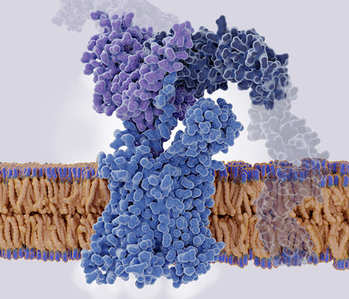
 Phosphatidic acid generation and its role in modulating negative membrane curvature (Bullen et al., 2016).
Regulation Mechanisms of Phosphatidic Acid in Biological Systems
Phosphatidic acid (PA) exerts its diverse biological effects through intricate regulatory mechanisms that govern its synthesis, degradation, and cellular localization within biological systems. Understanding these regulatory mechanisms is crucial for elucidating the physiological roles of PA and its implications in health and disease. In this section, we delve into the regulatory pathways that control PA metabolism and function in biological systems.
Biosynthesis Regulation
PA biosynthesis is tightly regulated to maintain cellular lipid homeostasis and meet the dynamic demands of membrane biogenesis and signaling processes. Regulation occurs at multiple levels, including transcriptional, post-transcriptional, and post-translational regulation of enzymes such as phosphatidate phosphatase (PAP) and diacylglycerol kinase (DGK). Cellular signaling pathways modulate the activity and expression of these enzymes in response to changing physiological conditions.
Degradation and Turnover
The turnover of PA molecules is governed by enzymatic degradation pathways that regulate cellular PA levels and signaling activity. Phosphatidic acid phosphatases (PAPs) catalyze the dephosphorylation of PA to generate diacylglycerol (DAG), while phospholipases such as phospholipase D (PLD) hydrolyze PA to produce phosphatidylglycerol (PG) and free fatty acids. Regulation involves feedback mechanisms, substrate availability, and cellular signaling cues that modulate enzyme activity and subcellular localization.
Subcellular Localization
Phosphatidic acid distribution within subcellular compartments is tightly regulated to facilitate its diverse biological functions and signaling activities. PA levels are dynamically regulated at membrane contact sites, where lipid biosynthesis and signaling processes converge. Membrane-associated proteins, lipid transporters, and organelle-specific signaling pathways regulate the localization and trafficking of PA molecules to distinct subcellular compartments.
Protein-Protein Interactions
Phosphatidic acid interacts with a myriad of protein targets to regulate their activity, localization, and function within cellular networks. These interactions modulate enzyme catalysis, protein conformational changes, and signal transduction pathways, shaping cellular responses to extracellular cues and environmental stimuli.
Crosstalk with Signaling Pathways
Phosphatidic acid integrates signals from cellular signaling pathways to coordinate cellular responses and adaptation to changing environmental conditions. PA serves as a node in signaling networks, where it intersects with protein kinase cascades, phospholipase pathways, and second messenger systems to amplify and propagate signaling events.
Phosphatidic Acid Analysis Methods
Accurate and reliable analysis of phosphatidic acid (PA) is essential for understanding its role in biological systems and its implications in health and disease. A variety of analytical techniques have been developed to quantify and characterize PA species in complex biological samples.
Liquid Chromatography-Mass Spectrometry (LC-MS)
Liquid chromatography-mass spectrometry (LC-MS) stands as a powerful technique for the analysis of phosphatidic acid due to its high sensitivity, resolution, and versatility. LC separates PA species based on their hydrophobicity and ionization properties, while mass spectrometry provides accurate mass measurements and structural information. By employing reverse-phase chromatography coupled with electrospray ionization (ESI) or atmospheric pressure chemical ionization (APCI), LC-MS enables the identification and quantification of PA species in biological samples with high throughput and sensitivity. Additionally, tandem mass spectrometry (MS/MS) techniques such as multiple reaction monitoring (MRM) allow for the selective detection of specific PA molecular species, enhancing analytical specificity and precision.
Gas Chromatography-Mass Spectrometry (GC-MS)
Gas chromatography-mass spectrometry (GC-MS) offers an alternative approach for the analysis of phosphatidic acid, particularly for the determination of fatty acid composition. GC separates PA-derived fatty acid methyl esters (FAMEs) based on their volatility and chromatographic retention times, followed by mass spectrometric detection for compound identification and quantification. By employing derivatization techniques such as methylation or silylation, GC-MS enables the analysis of free fatty acids released from PA molecules, providing insights into lipid metabolism and biosynthetic pathways. However, GC-MS requires extensive sample preparation and derivatization steps, limiting its applicability for the analysis of intact PA species and complex biological samples.
Select Services
Nuclear Magnetic Resonance (NMR) Spectroscopy
Nuclear magnetic resonance (NMR) spectroscopy serves as a valuable tool for the structural elucidation and quantitative analysis of phosphatidic acid in biological samples. NMR exploits the magnetic properties of atomic nuclei to probe molecular structure and dynamics, allowing for the identification of specific functional groups and chemical bonds within PA molecules. By acquiring one-dimensional (1D) and two-dimensional (2D) NMR spectra, including proton (1H) and phosphorus (31P) NMR, researchers can characterize PA species based on their chemical shifts, coupling patterns, and relaxation properties. Moreover, quantitative NMR techniques such as integration of peak areas or signal intensities enable the determination of PA concentrations in solution, offering insights into lipid metabolism and cellular physiology.
Thin-Layer Chromatography (TLC)
Thin-layer chromatography (TLC) remains a widely used technique for the qualitative analysis and separation of phosphatidic acid from other lipid species in biological samples. TLC relies on the differential migration of PA molecules on a solid support coated with a thin layer of stationary phase, followed by visualization and quantification of separated bands using staining reagents or fluorescence detection. While TLC provides rapid and cost-effective analysis of PA species, it lacks the sensitivity and specificity of modern chromatographic and spectroscopic techniques, limiting its utility for quantitative analysis and structural characterization.
Reference
- Bullen, Hayley E., and Dominique Soldati‐Favre. "A central role for phosphatidic acid as a lipid mediator of regulated exocytosis in apicomplexa." FEBS letters 590.15 (2016): 2469-2481.