Introduction to Exosomes
What Are Exosomes?
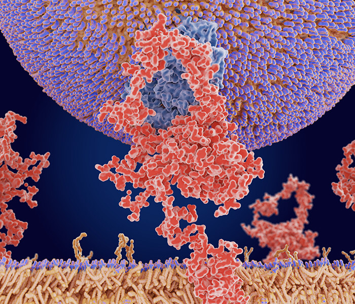
Exosomes, those tiny, membrane-bound vesicles secreted by cells, are much more than just cellular debris. They're carriers of a range of biomolecules—RNA, proteins, and lipids—that play pivotal roles in how cells communicate with one another. This ability to shuttle molecular information has made exosomes essential in unraveling disease mechanisms, particularly in cancer, and they're increasingly seen as key players in the future of precision medicine. One particularly intriguing aspect is the role of long non-coding RNAs (lncRNAs) within exosomes. Research involving over 9,000 tumor samples has shown that the expression levels of these lncRNAs correlate tightly with patient survival rates, marking them as potential biomarkers and therapeutic targets for cancer treatment. Interestingly, these non-coding RNAs are most active during the early stages of embryonic development, with their activity tapering off as the organism matures. This temporal pattern hints at a unique, specialized function during the early phases of life. All of this underscores the promise of exosome-based, personalized medical applications—an exciting prospect for the future of healthcare.
 Figure 1. Schematic representation of the different subtypes of extracellular vesicles (exosomes, microvesicles, and apoptotic bodies). (Marta Prieto-Vila et al,. 2021)
Role in Intercellular Communication
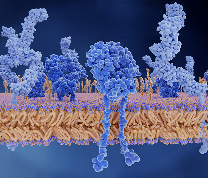
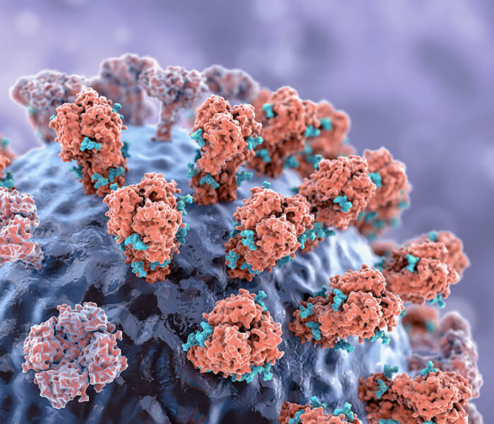
Exosomes are tiny vesicles that pack a powerful punch in the world of cellular communication. Acting like molecular couriers, they shuttle RNA, proteins, and lipids between cells, influencing a wide range of biological functions—from immune modulation to gene regulation. These microscopic messengers are becoming the focus of intense scientific research, not only because they play a crucial role in maintaining cellular homeostasis but also due to their potential as biomarkers for diseases and innovative therapeutic delivery systems.
But how exactly do scientists get their hands on these elusive exosomes? Isolating and purifying them from biological fluids like blood, urine, or saliva can be a tricky task. Let's take a look at some of the most commonly used techniques.
Exosome Isolation and Purification Techniques
Ultracentrifugation: The Classic Approach
Ultracentrifugation is often the first method that comes to mind when researchers think of isolating exosomes. It involves spinning the sample at incredibly high speeds, which generates a centrifugal force that separates particles based on their size and density. Larger particles, including exosomes, form a pellet at the bottom of the tube, making them easier to collect. While this technique is effective, it's not without its challenges. The process can be time-consuming and requires specialized equipment that might not always be available in every lab.
But for all its complexities, ultracentrifugation remains a gold standard, especially when researchers need to isolate exosomes from complex biological fluids with high efficiency. If you're looking for a deeper dive into exosome immunocapture protocols, you can explore our detailed guides and resources on this technique.
Size-Exclusion Chromatography (SEC): Size Matters
Imagine a column filled with tiny beads, each with pores that allow only certain molecules to pass through. This is the concept behind Size-Exclusion Chromatography (SEC). As a sample is passed through the column, larger particles, like exosomes, get caught in the larger pores and elute first, while smaller particles move through the column more slowly, exiting later. SEC offers excellent purity and is a great option for researchers working with large-scale applications. The method is particularly effective at separating exosomes from contaminants and other vesicles, ensuring high-quality isolation.
This method is a favorite for labs that need reproducibility and scalability without sacrificing purity. It's a reliable and efficient way to isolate exosomes when purity is key, and scalability is important.
Immunoaffinity Capture
For researchers looking for high specificity in exosome isolation, immunoaffinity capture might be the way to go. This technique uses antibodies designed to bind to specific markers present on the surface of exosomes. It's like using a high-tech magnet to pull out the exosomes from a pool of particles. The beauty of immunoaffinity capture lies in its specificity—researchers can isolate exosomes from a particular source or type of cell based on the antibodies they use.
However, while immunoaffinity capture is fantastic for obtaining high-purity exosome samples, there's a tradeoff in terms of yield. Because the technique targets only specific exosome populations, it may result in a lower overall yield compared to other methods like ultracentrifugation. Nonetheless, for studies focusing on particular biomarkers or vesicle populations, immunoaffinity capture is an invaluable tool. Check out our exosome proteomics services for more details on isolation and analysis.
Comparison of Methods
| Method | Yield | Purity | Processing Time | Cost | Scalability |
|---|---|---|---|---|---|
| Ultracentrifugation | Moderate | High | Long (>4 hours) | Low | Low |
| Size-Exclusion Chromatography | Moderate | High | Medium (1-2 hours) | Medium | Medium |
| Precipitation | High | Low | Short (<1 hour) | Low | High |
| Immunoaffinity Capture | Low | Very High | Medium (2-3 hours) | High | Low |
| Microfluidic Devices | Low | High | Very Short (<30 min) | High | Low |
Characterization Methods in Exosome Research
Exosome Characterization Techniques
Exosome characterization is crucial for understanding their biological role, isolating their contents, and exploring potential therapeutic applications. There are several advanced techniques used in exosome research, each offering unique advantages for analyzing their size, morphology, protein content, and functionality.
Nanoparticle Tracking Analysis (NTA)
Nanoparticle Tracking Analysis (NTA) is a widely used technique for determining the size distribution and concentration of exosomes. By tracking the Brownian motion of particles in a liquid suspension, NTA allows for the calculation of the size and concentration of nanoparticles in real-time, making it an essential tool for monitoring exosome release and dynamics.
Key advantages of NTA:
Real-time analysis: NTA provides immediate insights into the size distribution and concentration of exosomes, which is essential for understanding their release kinetics over time. This real-time capability is crucial for studies examining dynamic cellular processes, such as exosome secretion and uptake (Vogel et al., 2017).
Ideal for exosome size range: NTA is particularly effective for analyzing exosomes, which typically fall within the size range of 70-160 nm. NTA measures particles in this range with high precision, offering accurate and reproducible measurements of exosome populations (Lannigan et al., 2015).
Quantification of exosome release: NTA is widely used to quantify the release of exosomes from cells. It has been applied in studies to investigate exosome release under different conditions, such as stimulation by cytokines or in the context of disease (Pritchard et al., 2015).
For example, a study using NTA showed that unstimulated Jurkat and CEM T-cell lines released microvesicles in the size range of 70-90 nm (Lannigan et al., 2015).
Electron Microscopy (TEM)
Transmission Electron Microscopy (TEM) plays a crucial role in visualizing the morphology of exosomes, providing high-resolution imaging that reveals the characteristic cup-shaped appearance of exosomes.
Key features of TEM in exosome research:
- Sample preparation: TEM requires careful sample preparation, including fixation, dehydration, and embedding to preserve the exosome morphology. The procedure is labor-intensive but necessary for obtaining clear and reproducible results (Raposo et al., 1996).
- Negative staining: Negative staining enhances contrast and facilitates the visualization of exosome structures, especially for distinguishing between exosomes and other vesicles in complex biological samples (Crescitelli et al., 2013).
- Immuno-gold labeling: Immuno-gold labeling is a technique used in TEM to tag specific proteins on the surface of exosomes. This technique allows researchers to identify proteins of interest on exosome surfaces and confirm the presence of specific markers (Zhang et al., 2015).
TEM remains one of the most reliable methods for confirming the purity and morphology of isolated exosomes, ensuring that the vesicles under study are indeed exosomes and not other types of extracellular vesicles.
Western Blotting and Flow Cytometry
Western blotting and flow cytometry are widely employed techniques to identify and quantify specific proteins associated with exosomes. These methods provide valuable insights into the protein composition of exosomes, confirming their identity and assessing the purity of isolation methods.
Western Blotting: This technique is a powerful tool for protein identification and quantification. By detecting specific exosome markers such as CD9, CD63, and TSG101, western blotting allows researchers to confirm the presence of exosomes and assess the quality of exosome isolation protocols (Théry et al., 2006).
Flow Cytometry: Flow cytometry enables high-throughput analysis of exosome populations based on their surface markers. It is particularly useful for analyzing large numbers of samples and is often applied in clinical settings to analyze exosome populations in patient samples. Studies have shown that optimizing bead quantities and gel loading volumes can enhance the detection sensitivity of exosomes using flow cytometry (Im et al., 2014).
Mass Spectrometry in Exosome Proteomics
Mass spectrometry (MS) is a cornerstone for comprehensive proteomic analysis of exosomes. This technique enables the quantitative profiling of global proteomes and facilitates the identification of thousands of proteins in exosome samples, making it indispensable for proteomic research.
Key features of Mass Spectrometry in exosome research:
- Quantitative global proteome profiling: MS allows for detailed analysis of the entire proteome of exosomes, providing a comprehensive map of the proteins present in these vesicles. It has been used to identify a vast number of proteins that may be involved in disease processes, offering insights into potential therapeutic targets (Brahimi-Horn et al., 2011).
- Discovery of potential biomarkers: MS-based proteomics is instrumental in identifying novel biomarkers that can be used for disease diagnosis or therapeutic targeting. Several studies have demonstrated the application of MS in identifying biomarkers of cancer, cardiovascular disease, and neurodegenerative disorders in exosomes (Li et al., 2019).
 Figure 1. Different techniques used for isolation, characterization, and analysis of exosomes
Comparison of Exosome Characterization Techniques
| Technique | Key Advantages | Limitations | Typical Applications |
|---|---|---|---|
| Nanoparticle Tracking Analysis (NTA) | Real-time analysis, ideal for exosome size range (70-160 nm), quantifies exosome release | Can be affected by particle aggregation | Size distribution, concentration determination, monitoring exosome release |
| Electron Microscopy (TEM) | High-resolution imaging, confirms morphology, enhances contrast with negative staining | Requires extensive sample preparation | Visualizing exosome morphology, confirming purity |
| Western Blotting | Identifies specific proteins, provides quantification of exosome markers | Limited to known exosome markers, requires good antibodies | Identifying exosome-associated proteins (CD9, CD63, TSG101) |
| Flow Cytometry | High-throughput, analyzes surface markers, ideal for large sample volumes | May require optimization for exosome detection | High-throughput analysis of exosome populations based on surface markers |
| Mass Spectrometry (MS) | Comprehensive proteome profiling, identifies thousands of proteins, uncovers biomarkers | Expensive, requires highly specialized equipment and expertise | Proteomic analysis, biomarker discovery, therapeutic target identification |
Exosome Cargo Analysis
Exosome Proteomic Profiling
Proteomics is essential for analyzing the protein content of exosomes. This process helps identify key proteins involved in disease mechanisms and potential therapeutic targets.
Exosome RNA Sequencing
RNA sequencing allows for the study of exosome RNA content, offering insights into gene regulation and the role of exosomes in gene expression.
Exosome Lipidomic Studies
Lipidomics provides an analysis of the lipid content in exosomes. This is crucial for understanding membrane properties and the role of lipids in cellular communication.
Select Service
Exosome Proteomics
Oerview of Proteomic Profiling in Exosomes
Proteomic profiling involves the identification and quantification of proteins found in exosomes. The molecular composition of these proteins provides insights into the biological roles of exosomes in disease and health. Explore our exosome proteomics services for detailed analysis.
Exosome Mass Spectrometry: Advanced Analytical Techniques
Mass spectrometry for exosome research provides a deeper understanding of the protein cargo inside exosomes. High-resolution MS techniques allow for detailed characterization, revealing low-abundance proteins that might otherwise go undetected. With the increasing use of mass spectrometry in exosome analysis, the potential for biomarker discovery and targeted therapies is growing rapidly.
Applications of Exosome Research
Diagnostic Biomarkers
Exosomes are increasingly being recognized for their potential in liquid biopsy-based diagnostics. They carry disease-specific markers that can be used to detect early-stage diseases such as cancer and neurodegenerative disorders.
Therapeutic Delivery Systems
Exosomes are naturally suited for drug delivery due to their ability to transport proteins, RNA, and small molecules. Their biocompatibility and low immunogenicity make them ideal candidates for targeted therapeutic delivery.
Role in Disease Pathogenesis
Exosomes play a significant role in the progression of various diseases. In cancer, for example, they can transfer oncogenic proteins and RNAs between tumor cells, promoting metastasis. Their involvement in neurological diseases and immune response further emphasizes their importance in understanding disease mechanisms.
Challenges and Future Directions in Exosome Technology
Standardization of Isolation Methods
One of the main challenges in exosome research is the lack of standardized isolation techniques. Different methods can lead to variations in exosome purity and yield, which can impact the reproducibility of research findings.
Scalability for Clinical Applications
For exosome-based therapies to be widely used in clinical settings, methods for large-scale isolation and purification must be developed. This includes addressing issues related to the scalability of exosome isolation techniques.
Regulatory Considerations
As exosome-based therapeutics enter clinical trials, regulatory frameworks will need to be established to ensure their safety and efficacy. Standardized guidelines for clinical use will be essential to gain regulatory approval.
Emerging Technologies
Recent advancements in microfluidic devices, CRISPR, and machine learning algorithms are revolutionizing exosome research. These technologies hold the potential to improve the accuracy, speed, and scalability of exosome analysis.
Real Data and Industry Statistics
| Metric | Value | Source |
|---|---|---|
| Global Exosome Market Size (2024) | USD 1.4 Billion | Grand View Research (2021) |
| Projected CAGR of Exosome Market (2024-2030) | 18.7% | Grand View Research (2021) |
| Number of Exosome Proteins Identified in Research | Over 1,000 | Journal of Proteome Research, 2020 |
| Exosome-Based Therapeutic Applications (2022) | 50+ Clinical Trials Ongoing | ClinicalTrials.gov (2022) |
Conclusion
Exosome research has become one of the most exciting fields in molecular biology. With advanced techniques like mass spectrometry and nanoparticle tracking analysis, scientists are uncovering new possibilities for diagnostics, therapeutic delivery, and disease treatment. However, there are still challenges to overcome, such as the standardization of isolation methods and scalability for clinical applications.
At Creative Proteomics, we provide comprehensive exosome analysis services, from isolation to proteomics, lipidomics, and RNA sequencing. Our cutting-edge technologies and expertise can help you unlock the full potential of exosome research for your studies or clinical applications.
Contact us today to learn more about how our exosome analysis services can advance your research.
References
- Vogel, R., F. T. Wilhelm, R. et al. (2017). "NTA in cellular biology: Current applications in nanomaterials and nanobiotechnology." Journal of Nanobiotechnology, 15(1), 12.
- Lannigan, J., et al. (2015). "Size-dependent characteristics of exosomes and microvesicles released by human T-cells." Journal of Extracellular Vesicles, 4(1), 29075.
- Pritchard, D., et al. (2015). "Tracking exosome release and uptake in immunological processes." Journal of Immunological Methods, 428, 30-37.
- Crescitelli, R., et al. (2013). "Distinct RNA profiles in subpopulations of extracellular vesicles: apoptotic bodies, microvesicles and exosomes." Journal of Extracellular Vesicles, 2(1), 20677.
- Théry, C., et al. (2006). "Isolation and characterization of exosomes from cell culture supernatants and biological fluids." Current Protocols in Cell Biology, 30, 3.22.1–3.22.29.