Introduction to Exosome Proteomics
Exosome proteomics is revolutionizing our understanding of cell-to-cell communication, disease progression, and the development of targeted therapies. These nanoscale vesicles, once regarded simply as byproducts of cellular processes, are now recognized as critical carriers of molecular information. Through the analysis of protein profiles in exosomes, researchers are identifying novel biomarkers for early disease detection, advancing therapeutic strategies, and even engineering exosomes for targeted drug delivery.
What distinguishes exosome proteomics as a transformative force in contemporary medicine? This paper will examine the most recent developments, practical applications, and ongoing challenges within this dynamic field.
As exosome proteomics continues to bridge the gap between fundamental research and clinical implementation, its capacity to reshape diagnostic and therapeutic paradigms is only beginning to emerge.
What Are Exosomes? Please refer to Exosome Analysis: Cutting-Edge Research Technology & Proteomic Profiling.
Why Study the Proteomics of Exosomes?
Role in Intercellular Communication
Exosomal proteins play a pivotal role in intercellular communication, allowing cells to exchange crucial signals. These proteins help regulate immune responses, tissue repair, and even cancer progression. By carrying proteins, lipids, and genetic material from one cell to another, exosomes help cells communicate in a way that can influence entire biological systems.
Applications of Exosome Proteomics
Exosome proteomics has indeed emerged as a transformative tool in cancer research, offering significant advancements in early detection, monitoring, and personalized medicine. Recent studies have demonstrated the power of exosome-derived protein biomarkers in revolutionizing cancer diagnostics and treatment strategies.
Early Detection and Diagnosis
Exosomes exhibit a distinct advantage in cancer diagnosis, as they harbor specific signatures reflective of the tumor's genetic and proteomic profile. A study by Goel et al. (2024) demonstrated the potential of an exosome-based liquid biopsy for early detection of pancreatic cancer. When combined with the CA19-9 biomarker, their approach accurately detected 97% of stage 1-2 pancreatic cancers. This high accuracy in early-stage detection highlights the potential of exosome proteomics to significantly improve cancer diagnosis before tumor progression.
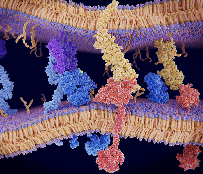
 The role of exosomal proteins in cancer biology. (Xinyi Wang et al,. 2022)
Liquid Biopsies and Non-invasive Monitoring
Exosome proteomics offers a non-invasive alternative to traditional tissue biopsies, enabling the use of liquid biopsies for cancer detection and monitoring. A study by Xu et al. (2024) developed a novel computational approach using a random forest classifier to define exosome protein panels that serve as effective biomarkers for plasma, serum, or urine across cancer types. This method demonstrated high accuracy in differentiating between cancer types, further enhancing the diagnostic value of exosome-based approaches.
Prognosis and Personalized Medicine
Exosomal protein profiles have shown promise in predicting disease progression, treatment response, and recurrence. A study analyzing proteomics data from plasma or serum-derived exosomes from patients with five common cancer types (breast, colorectal, glioma, lung, and pancreatic) demonstrated that a set of five proteins could reliably differentiate between these cancer types with high accuracy. This exosome protein-based classification model can enhance diagnostic value and potentially guide personalized treatment strategies.
Biomarker Discovery for Diseases
Exosome proteomics has significantly advanced biomarker discovery across various diseases, particularly cancers and neurodegenerative diseases. A review by Goel et al. (2024) highlighted the potential of exosomes as reliable biomarkers for cancer detection and diagnosing specific cancer types. This research underscores the growing importance of exosome-based diagnostics in clinical settings.
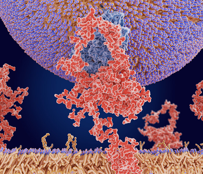
 Proteomic profile of extracellular vesicles (Ayuko Hoshino et al,. 2020)
Therapeutic and Diagnostic Implications
Exosomes are gaining recognition as both diagnostic and therapeutic agents, with significant applications in drug delivery systems and non-invasive diagnostics. A comprehensive review by Zhang et al. (2020) described the advantages of exosomes as an effective liquid biopsy tool and the progression of exosome extraction methods. This research emphasizes the potential of exosomes in advancing both diagnostic and therapeutic approaches in cancer management.In conclusion, exosome proteomics represents a promising frontier in cancer research and diagnosis, offering new possibilities for early detection, monitoring, and personalized treatment strategies. As our understanding of exosome biology and their role in cancer continues to grow, we can expect further advancements that will shape the future of oncology and improve patient outcomes.
Learn more about exosome-based diagnostic techniques here: Exosome Analysis & Profiling.
Explore Creative Proteomics' exosome-related services: Exosome Proteomics Services.
Technologies Used in Exosome Proteomics
The study of exosome proteomics has been greatly enhanced by the development of advanced technologies that enable precise identification, quantification, and analysis of the proteins contained within exosomes. These technologies are crucial for unraveling the complex roles exosomes play in intercellular communication, disease progression, and potential therapeutic applications. Below are the prominent technologies and emerging methods used in exosome proteomics:
Mass Spectrometry (MS)-Based Techniques
Liquid Chromatography-Tandem Mass Spectrometry (LC-MS/MS):
Widely used for high-resolution analysis of exosomal proteins, LC-MS/MS enables deep profiling of the exosomal proteome. For example, nano-ESI–MS/MS has been applied to detect thousands of proteins from minimal sample volumes, making it ideal for limited exosome yields.
LC-MS/MS Application in Exosome Proteomics:
- Identification and Quantification: MS enables precise identification and quantification of proteins present in exosomes, even at low abundances.
- High Sensitivity: Essential for analyzing the minute quantities of proteins in small exosome samples.
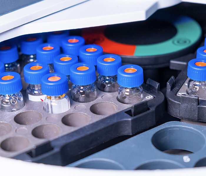
 Exosomal proteomic profile Experimental workflow by LC-MS/MS
Trapped Ion Mobility Spectrometry-Mass Spectrometry (TIMS-MS):
TIMS-MS has enabled significant advancements in exosome proteomics, including biomarker discovery and understanding disease mechanisms. Its ability to provide deep proteome coverage from small sample volumes makes it a powerful tool for exosome research and potential clinical applications.
Key features and benefits of TIMS-MS include:
- Enhanced Sensitivity and Speed: TIMS-MS combines ion mobility spectrometry with mass spectrometry, providing enhanced sensitivity and faster analysis times, which is crucial for analyzing small exosomal samples.
- High Reproducibility: This technique ensures consistent results across experiments, which is important for reliable data.
- Deep Proteome Coverage: TIMS-MS allows the identification of thousands of proteins even from minimal sample inputs.
- High Reproducibility: TIMS-MS ensures consistent results across experiments, which is crucial for reliable data in exosome research.
- Improved Separation: The addition of ion mobility separation reduces spectral complexity, leading to better protein identification.
- Applicability to Small Sample Volumes: TIMS-MS is particularly useful for analyzing the minute quantities of proteins in small exosome samples, making it ideal for clinical applications.
Data-Independent Acquisition (DIA) and 4D Proteomics
The combination of DIA (Data-Independent Acquisition) with 4D proteomics represents a new frontier in exosome analysis. These techniques allow for the comprehensive analysis of all proteins in a sample, providing a deeper understanding of exosome composition and enabling the discovery of new biomarkers for diseases.
Data-Independent Acquisition (DIA)
DIA is a mass spectrometry approach where all ions in a sample are fragmented and analyzed simultaneously. This method:
- Captures data on all detectable peptides, increasing the identification of low-abundance proteins.
- Provides reliable quantification across multiple samples and conditions, ensuring consistent data output for proteomics analyses.
4D Proteomics
4D proteomics incorporates an additional dimension of separation, typically using ion mobility:
- Reduces spectral complexity and improves protein identification by separating ions based on their shape and mobility before mass analysis, leading to enhanced resolution.
- Enhances separation capacity, leading to deeper proteome coverage by reducing interference from complex mixtures.
Synergistic Effects of DIA and 4D Proteomics
When combined, DIA and 4D proteomics offer significant benefits for exosome analysis:
- Deeper proteome coverage and more accurate quantification, enabling the identification of proteins present in lower quantities.
- Improved sensitivity and detection of low-abundance proteins, crucial for identifying subtle biological markers and pathways.
DIA and 4D Proteomics Applications in Exosome Proteomics
- In-Depth Protein Profiling:
DIA and 4D proteomics together allow the identification of a broader range of exosomal proteins, including those present in low abundances, which is critical for accurate biomarker discovery. - Biomarker Discovery:
Facilitates the identification of novel disease biomarkers, which can aid in the development of targeted therapies. - Disease Mechanism Elucidation:
Helps understand complex protein interactions and pathways involved in disease progression, offering insights into potential therapeutic targets.
DIA and 4D Proteomic Recent Advancements
- 4D-DIA Quantitative Proteomics:
Based on the timsTOFPro mass spectrometer, 4D-DIA offers faster and more sensitive scanning compared to traditional 3D-DIA technologies, achieving comprehensive improvements in proteomics coverage, sensitivity, and throughput. - MaxDIA Software:
A platform for analyzing DIA proteomics data within the MaxQuant software environment, which significantly enhances deep proteome coverage and protein quantification reliability. - Application in Clinical Studies:
A recent study on obstetric antiphospholipid syndrome (OAPS) used 4D-DIA-MS to identify 4270 and 3328 proteins from large and small extracellular vesicle (EV) enriched fractions, respectively, showcasing the potential of DIA-4D methods in clinical biomarker discovery.
Explore innovative exosome isolation techniques with Creative Proteomics: PEG-based Precipitation Protocol for Exosome Isolation.
Select Service
Innovative Exosome Isolation Techniques
The efficiency of exosome isolation is a critical determinant of the accuracy and reliability of downstream proteomic analyses. The quality and purity of exosome preparations directly influence experimental outcomes, making the choice of isolation method crucial. Traditional approaches, such as ultracentrifugation, while widely used, are time-consuming and may not yield sufficiently pure exosome preparations due to contamination with other extracellular vesicles or cellular debris. This section explores recent advancements in exosome isolation techniques, including Polyethylene Glycol (PEG)-based precipitation, Size-Exclusion Chromatography (SEC), and immunoaffinity-based methods.
Polyethylene Glycol (PEG)-Based Precipitation Protocols
Principle: Polyethylene glycol (PEG) is employed to precipitate exosomes by decreasing their solubility, facilitating their collection through centrifugation at lower speeds.
Advantages:
Simplicity and Efficiency: PEG precipitation is a straightforward and rapid technique, often more time-efficient than ultracentrifugation. Its reduced technical complexity makes it an attractive option for laboratories with limited resources. Additionally, it significantly shortens preparation time.
High Yield: This method has proven effective in isolating exosomes from large sample volumes, maintaining high recovery rates. For instance, PEG precipitation has successfully isolated exosomes from plasma and serum samples, achieving substantial yields with minimal loss.
Applications:
Clinical Samples: PEG is particularly beneficial for isolating exosomes from biological fluids such as plasma or urine, where exosome concentrations tend to be low. Its speed and efficiency make it suitable for clinical settings where sample volume and time constraints are significant.
Considerations:
Purity: A primary limitation of PEG precipitation is the potential for co-precipitation of other extracellular vesicles or proteins, complicating subsequent analyses. Additional purification steps, such as ultracentrifugation or immunoaffinity capture, may be necessary to enhance purity.
Size-Exclusion Chromatography (SEC)
Mechanism: SEC separates particles based on size, allowing for the isolation of exosomes while excluding larger particles and protein complexes. Exosomes, being smaller than cellular debris, elute faster through the column, whereas larger particles are retained.
Benefits:
High Purity: SEC is renowned for its ability to yield highly purified exosome fractions. Unlike PEG precipitation, which can result in co-precipitation, SEC produces cleaner exosome isolates, making it ideal for sensitive proteomic analyses.
Non-Damaging: Unlike ultracentrifugation, SEC does not expose exosomes to harsh physical conditions (e.g., high centrifugal forces), thus preserving vesicle integrity.
Immunoaffinity-Based Isolation
Approach: This technique leverages antibodies targeting specific exosomal surface markers, such as CD9, CD63, and CD81, to selectively capture exosomes. These antibodies are often conjugated to magnetic beads or other solid-phase substrates, facilitating rapid isolation using a magnetic field or centrifugation.
Advantages:
Specificity: Immunoaffinity-based isolation offers exceptional specificity, enabling the selective capture of exosomes from specific cell types or those bearing particular biomarkers. This is especially useful when studying disease-associated exosomes, such as those involved in cancer, where specific surface markers are linked to tumor-derived vesicles.
High Sensitivity: This method allows for the isolation of exosomes from low-concentration clinical samples, such as serum or urine, where exosome quantities may be limited.
Limitations:
Cost and Complexity: Immunoaffinity isolation can be more expensive and labor-intensive than methods like PEG precipitation or SEC. The need for specific antibodies for targeted exosomal markers increases both cost and procedural complexity.
Emerging Isolation Methods
Microfluidic Devices: Microfluidics represents a promising new approach to exosome isolation. These devices manipulate small volumes of fluid through precise channels, enabling efficient exosome capture from complex samples. They offer rapid isolation with minimal sample volume, making them particularly advantageous for clinical diagnostics.
Ultrafiltration and Dialysis: These techniques utilize membrane filters to separate exosomes based on size. While simpler than SEC, they can process larger volumes of sample, although they may offer lower purity.
By adopting these advanced isolation techniques, researchers can achieve higher yields and enhanced purity, facilitating more accurate proteomic profiling and improving disease diagnosis and treatment. However, each method has inherent advantages and limitations, and the selection of the appropriate technique often depends on the specific research objectives and available resources.
Emerging Methods in Exosome Proteomics
Photocleavable Surfactants:
Concept: Surfactants, commonly used for protein solubilization, can interfere with mass spectrometry (MS) analysis. Photocleavable surfactants, which degrade upon exposure to light, can be employed after protein extraction to eliminate such interference, thereby improving the quality of MS data.
Advantages:
Enhanced Protein Solubilization: These surfactants improve the recovery of membrane proteins from exosomes, enhancing the identification of membrane-associated biomarkers.
Simplified Workflow: Photocleavable surfactants streamline the MS analysis process by reducing the need for extensive cleanup steps, thereby saving time and resources.
Impact on Exosome Proteomics:
Increased Throughput: The use of photocleavable surfactants accelerates sample preparation, allowing for the processing of larger sample numbers, which is crucial for large-scale exosome proteomics studies.
Improved Data Quality: These surfactants minimize the presence of contaminants that may hinder protein identification and quantification in exosome proteomics.
Microfluidics and Lab-on-a-Chip Technologies:
Overview: Microfluidic devices integrate multiple laboratory functions on a single chip, offering a highly efficient platform for exosome analysis.
Benefits in Exosome Analysis:
Rapid Isolation and Analysis: Microfluidic systems significantly reduce processing times for exosome isolation and analysis, enabling faster and more efficient separation techniques.
Reduced Sample Volumes: These technologies are particularly useful in clinical settings, where sample volumes are often limited.
Applications:
Point-of-Care Testing: Microfluidic devices have the potential to facilitate bedside exosome analysis, providing real-time diagnostics and disease monitoring.
Challenges:
Device Standardization: Despite their promise, the widespread adoption of microfluidic devices is hindered by challenges related to consistent fabrication and performance, making standardization critical for reliable clinical use.
Advancements in Bioinformatics Tools:
Data Analysis and Interpretation: The large datasets generated by high-throughput proteomic techniques require advanced bioinformatics tools for effective processing and interpretation.
Handling Large Datasets: As exosome proteomics generates vast amounts of data, bioinformatics methods such as machine learning and data mining are increasingly used to uncover meaningful patterns from complex datasets.
Integration with Other Omics Data:
Holistic Understanding: Integrating proteomic data with genomics and metabolomics provides a more comprehensive understanding of disease mechanisms and potential therapeutic targets.
These technological advancements in exosome isolation and analysis are poised to enhance the capabilities of proteomics, enabling more accurate and efficient disease diagnosis, as well as the development of novel therapeutic approaches.
Summary Of Exosome Proteomics Technologies
| Technology | Overview | Applications in Exosome Proteomics | Advantages | Additional Notes |
|---|---|---|---|---|
| Mass Spectrometry (MS) | Technique for measuring mass-to-charge ratio of ions. | - Identification and quantification of proteins in exosomes, even at low abundance. | - High sensitivity, essential for analyzing small exosome samples. | - Crucial for proteomics analysis. |
| Trapped Ion Mobility Spectrometry-Mass Spectrometry (TIMS-MS) | Combines ion mobility spectrometry with MS for enhanced sensitivity. | - High reproducibility and deep proteome coverage. | - Faster analysis and enhanced sensitivity. | - Useful for biomarker discovery and disease mechanism understanding. |
| Data-Independent Acquisition (DIA) | Mass spectrometry approach where all ions are fragmented and analyzed simultaneously. | - Comprehensive analysis of all peptides, increasing chances of identifying low-abundance proteins. | - Reliable quantification and higher coverage across samples and conditions. | - Increases data capture for low-abundance proteins. |
| 4D Proteomics | Adds a dimension of separation (e.g., ion mobility) to standard proteomics. | - Identifies a broader range of exosomal proteins. | - Enhanced separation, reduced spectral complexity, improved protein identification. | - Combines well with DIA for deeper proteome coverage. |
| Polyethylene Glycol (PEG)-Based Precipitation | PEG precipitates exosomes by reducing their solubility. | - Isolates exosomes from large volumes with high yield, useful for clinical samples. | - Simple, efficient, and faster compared to ultracentrifugation. | - May require additional purification for purity. |
| Size-Exclusion Chromatography (SEC) | Separates particles based on size. | - High purity exosome preparations for sensitive proteomic analysis. | - Provides cleaner exosome preparations than other methods. | - Ideal for isolating high-purity exosomes. |
| Immunoaffinity-Based Isolation | Uses antibodies to capture exosomes based on specific surface markers. | - Selective capture of exosomes from specific cell types. | - High specificity for exosomal isolation. | - Expensive and labor-intensive. |
| Microfluidic Devices | Miniaturized devices for rapid exosome isolation. | - Efficient for processing small sample volumes in clinical settings. | - Reduces processing time and sample volume requirements. | - Still needs standardization for widespread use. |
| Ultrafiltration and Dialysis | Utilizes membrane filters to separate exosomes based on size. | - Efficient separation based on size for exosome isolation. | - Simple technique with low-cost setup. | - Effective for isolating exosomes with minimal sample loss. |
| Photocleavable Surfactants | Surfactants that can be degraded by light after protein extraction to eliminate interference. | - Improves recovery of membrane proteins from exosomes. | - Enhanced protein solubilization and reduced interference in MS analysis. | - Increases throughput and improves data quality. |
| Microfluidics and Lab-on-a-Chip | Devices that integrate laboratory functions on a single chip. | - Allows rapid isolation and analysis of exosomes from limited clinical sample volumes. | - Reduced sample volumes, ideal for clinical applications. | - Requires consistent fabrication for reliability. |
| Advancements in Bioinformatics Tools | New tools to process and interpret data generated by high-throughput proteomics. | - Integration of proteomic data with genomics and metabolomics for comprehensive biological insights. | - Enables handling large datasets for holistic understanding. | - Important for large-scale proteomic data analysis. |
Challenges in Exosome Proteomics
While exosome proteomics holds great promise, it also faces several challenges:
Technical difficulties in isolation and characterization: Exosomes are small and heterogeneous, making them difficult to isolate and study with precision.
Standardization issues: There are currently no universally accepted protocols for exosome isolation or proteomics analysis, which can lead to inconsistent results across different studies.
Reproducibility concerns: The lack of standardized methods means that reproducibility across laboratories remains a significant issue.
To overcome these challenges, Creative Proteomics provides comprehensive protocols and customized solutions for exosome isolation and analysis.
Integration with Multi-Omics Approaches
The integration of exosome proteomics with other "omics" technologies, such as genomics, transcriptomics, and metabolomics, is poised to revolutionize the field. This multi-omics approach will provide a comprehensive understanding of the molecular mechanisms underlying health and disease. For example, advanced mass spectrometry (MS)-based proteomics has been successfully combined with transcriptomic and metabolomic data to identify novel biomarkers for cancer diagnosis and prognosis, as demonstrated in studies on prostate and bladder cancers (Wang et al., 2023). Such integration enables deeper insights into disease progression and therapeutic targets.
Machine Learning for Biomarker Discovery
Machine learning (ML) is transforming biomarker discovery by enabling the analysis of large-scale proteomics datasets. Recent studies have shown that ML algorithms, such as random forest classifiers, can identify exosome protein panels with high accuracy for early cancer detection and classification. For instance, a study identified five exosome proteins that reliably distinguished cancer exosomes from non-cancer exosomes across five common cancer types using plasma, serum, or urine samples (Xu et al., 2024). This approach demonstrated excellent sensitivity and specificity, paving the way for scalable and non-invasive diagnostic methods.
Challenges and Future Prospects
Despite these advancements, challenges remain in standardizing exosome isolation methods to ensure reproducibility and high yield for large cohorts. Current techniques like ultracentrifugation are time-consuming and may result in low protein recovery. Future innovations in isolation technologies will be critical for advancing exosome-based diagnostics (Wang et al., 2023). Additionally, the integration of ML with multi-omics data holds promise for identifying disease-specific biomarkers with unprecedented precision.
Conclusion: The Future of Exosome Proteomics
Exosome proteomics is transforming our comprehension of cellular communication and unlocking new possibilities for diagnostics and therapeutic applications. As technological advancements, particularly in mass spectrometry and artificial intelligence, continue to progress, this field is set to make significant contributions to a wide range of medical areas, including cancer detection and personalized medicine.
Explore Creative Proteomics' full range of services: Exosome Proteomics Solutions.
References
- Xu, Y., Ku, X., Wu, C., Cai, C., Tang, J., & Yan, W. (2024). A novel machine learning algorithm selects proteome signature to detect cancer and classify cancer types using exosomes. eLife, 13, e90390. https://doi.org/10.7554/eLife.90390
- Goel, A., Xu, Y., & Yin, J. (2024). An Exosome-based Liquid Biopsy Shows Promise for Early Detection of Pancreatic Cancer. American Association for Cancer Research (AACR) Annual Meeting 2024. https://www.aacr.org/about-the-aacr/newsroom/news-releases/an-exosome-based-liquid-biopsy-shows-promise-for-early-detection-of-pancreatic-cancer/
- Goel, A., & Chauhan, S. S. (2022). Exosomes and cancer - Diagnostic and prognostic biomarkers and therapeutic implications. Signal Transduction and Targeted Therapy, 7(1), 298. https://doi.org/10.1038/s41392-022-01133-5
- Zhang, Y., Liu, Y., Liu, H., & Tang, W. H. (2020). Application of exosomes as liquid biopsy in clinical diagnosis. Signal Transduction and Targeted Therapy, 5(1), 144. https://doi.org/10.1038/s41392-020-00258-9
- Sinha, D., Roy, S., Saha, P., Chatterjee, N., & Bishayee, A. (2024). Exosomes: a promising avenue for cancer diagnosis beyond treatment. Cell Death Discovery, 10(1), 12. https://doi.org/10.1038/s41420-023-01710-9
- Wang, Y., Ku, X., Wu, C., Cai, C., Tang, J., & Yan, W. (2023). Proteomic dissection of exosome cargo: Progress and future directions. Frontiers in Oncology, 13(1), Article 144. Xu, Y., Ku, X., Wu, C., Cai, C., Tang, J., & Yan, W. (2024). A novel machine learning algorithm selects proteome signature to detect cancer and classify cancer types using exosomes. eLife, 13(1), Article e90390. https://doi.org/10.7554/eLife.90390
- Wang, X., Chen, H., & Yuan, G.-C. (2020). Proteomic analysis of extracellular vesicles: Advances and challenges in biomarker discovery. Genome Biology, 21(1), Article 102.