Western Blot Detection Service
Creative Proteomics offers Western Blot Detection Services and western blot analysis.We specialize in protein extraction from tissues or cells, Western blot construction, immunostaining with customized antibodies, and quantitative analysis.Our services include high-quality digital imaging to support your research needs.
Submit Your Request Now
×- What Is Western Blot
- How It Works
- Our WB Detection Service
- Applications
- Our Advantages
- Sample Requirements
- FAQs
- Demo
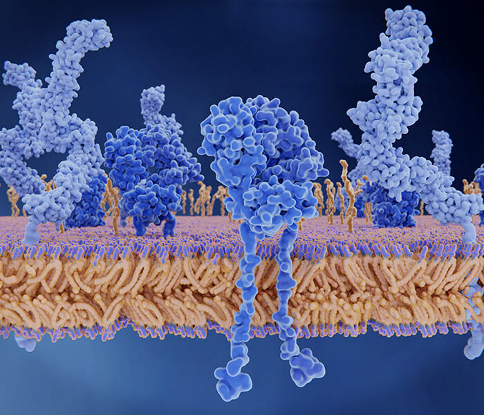
What Is Western Blot?
Western blotting, or immunoblotting, first introduced by Towbin et al. in 1979, has become an essential technique for studying protein expression. Over the past 40 years, Western blotting has become one of the most commonly employed tools in protein research. This method relies on antigen-antibody interactions, employing specific antibodies to identify and analyze target proteins. It allows researchers to perform both qualitative assessments and semi-quantitative evaluations of the protein of interest.
How Dose Western Blot Work?
The Principle of Western Blot
The principle of Western Blot is based on the specific interaction between antigens and antibodies. First, proteins in the sample are separated by polyacrylamide gel electrophoresis (SDS-PAGE) according to their molecular weight. The separated proteins are then transferred onto a solid-phase support membrane, such as a nitrocellulose or PVDF membrane, through electrophoretic transfer. Specific antibodies are then used to bind to the target protein. Detection and analysis of the target protein's expression are achieved by introducing a labeled secondary antibody and a subsequent colorimetric or chemiluminescent reaction, enabling both qualitative and semi-quantitative analysis.
 Figure 1. The workflow of western blot assay.(Meftahi, Biochem Mol Biol Educ, 2021)
Steps of Western Blot
Step 1. Sample Preparation:
Proteins are extracted from cells or tissues using lysis buffers containing detergents and inhibitors to prevent degradation. The protein concentration is quantified (e.g., using Bradford assay), and the samples may undergo cleanup to remove interfering substances like nucleic acids or salts.
Step 2. Gel Electrophoresis:
Proteins are separated by size using SDS-PAGE. The samples are denatured with heat and reducing agents before loading onto the gel, and the gel matrix and acrylamide concentration are optimized for the best resolution of the target proteins.
Step 3.Transfer of proteins:
After separation, proteins are transferred from the gel to a membrane (such as nitrocellulose or PVDF) through electroblotting. This step immobilizes the proteins on the membrane for subsequent detection.
Step 4. Antibody Blotting:
The membrane is blocked to prevent nonspecific binding using blocking agents like BSA or non-fat milk. A primary antibody specific to the target protein binds to the immobilized protein, followed by a labeled secondary antibody that amplifies the signal for detection.
Step 5. Detection:
Various detection methods, including chemiluminescence, fluorescence, or colorimetric systems, are used. Enzymatic methods (like HRP-based chemiluminescence) or fluorophores conjugated to antibodies generate measurable signals.
Step 6. Visualization:
The signals are captured using X-ray film, CCD cameras, or laser scanners. Fluorescent signals are directly detected using appropriate excitation and emission filters.
Step 7. Data Analysis:
Protein bands are analyzed based on their intensity and position relative to molecular weight markers. Quantitative comparisons are made by measuring band intensity and normalizing against loading controls or total protein stains.
Advantages of Western Blot
- Widely used for protein expression level detection.
- Identification of specific proteins; can also be used for quantitative and semi-quantitative analysis of target proteins.
- Suitable for subsequent analysis of protein-protein, protein-DNA, and protein-RNA interactions.
Issues and Strategies Frequently Encountered in Western Blot Experiments
| Experimental Issue | Possible Reasons | Recommended Solutions |
|---|---|---|
| Absence of Target Band | Low expression of target protein | * Increase sample loading, concentrate target protein, use more sensitive reagents |
| Insufficient antibody potency | * Use freshly prepared primary antibody, avoid repeated freeze-thaw cycles; increase primary or secondary antibody concentration, extend incubation time | |
| Low transmembrane efficiency | * Choose appropriate transfer method, improve transfer efficiency | |
| Non-parallel, Curved Bands | Rapid electrophoretic migration or high migration temperature | * Adjust electrophoresis settings such as pH, voltage, etc., decrease ambient temperature during electrophoresis |
| High Background | Inappropriate blocking conditions | * Extend blocking time or change blocking agent |
| High antibody concentration | * Reduce primary/secondary antibody concentration and extend incubation time | |
| Cross-reaction between antibody and blocking agent | * Use additives like Tween 20 and detergents to minimize cross-reaction |
Our Western Blot Detection Service
Creative Proteomics provides western blotting analysis for the detection of a specific target protein out of a complex protein mixture, e. g. tissue homogenate or cell extract, using highly selective and sensitive antibody-antigen interactions.
We offer the following Western Blot Service, but not limited to:
- Protein Extraction: Extraction of protein samples from human or animal cells, tissues, bacteria, etc.
- Protein Quantification: Semi-quantitative analysis of proteins in the samples.
- Western Blot Detection
(1) Electrophoresis: Separation of protein samples using SDS-PAGE.
(2) Wet Transfer.
(3) Hybridization: Identification of specific proteins on the membrane using antibodies.
(4) Visualization: Fluorescence staining, black bands on a white background image - Internal Reference Protein Detection
- Pre-experiment and Formal Experiment Services.
Applications of Western Blot Detection
Detection of Protein Isoforms
Detects different protein isoforms by targeting specific epitopes, identifying variations with minimal size differences as biomarkers.
Detection of Protein-DNA Interactions
Southwestern blotting identifies DNA-binding proteins and transcription factors by using radiolabeled DNA probes to detect interactions.
Detection of Post-Translational Modifications (PTMs)
Used to detect and characterize PTMs, particularly for low-abundance, endogenous modifications with specific antibodies.
Antibody Development and Epitope Mapping
Ideal for developing and identifying antibodies, as well as mapping specific epitopes by probing membranes with antibodies.
Our Advantages
- Utilization of different protein concentration determination methods (Lowry or BCA) for various protein samples.
- Ensuring experiment quality through rigorous QC (Quality Control): Accurate quantification using internal references, with quantification values showing less than 8% variation across multiple experiments on the same sample.
- Ensuring result accuracy.
- Comprehensive technical services, ranging from gene synthesis, cell transfection, vector construction, protein expression and purification, to protein functional studies.
- Years of antibody research experience, with extensive knowledge in antibody selection to mitigate false-positive results at the source.
Sample Requirements
| Sample type | Recommended sample size | |
|---|---|---|
| Animal tissues | Hard tissues (bones, hair) | 300-500mg |
| Soft tissues (leaves, flowers of woody plants, herbaceous plants, algae, ferns) | 200mg | |
| Plant tissues | Hard tissues (roots, bark, branches, seeds, etc.) | 3-5g |
| Microbes | Common bacteria, fungal cells (cell pellets) | 100μL |
| cells | Suspension/adherent cultured cells (cell count/pellet) | >1*107 |
| Fluids | Plasma/serum/cerebrospinal fluid (without depletion of high abundance proteins) | 20μL |
| Plasma/serum/cerebrospinal fluid (with depletion of high abundance proteins) | 100μL | |
| Follicular fluid | 200μL | |
| Lymph, synovial fluid, puncture fluid, ascites | 5mL | |
| Others | Saliva/tears/milk | 3-5mL |
| Culture supernatant (serum-free medium cannot be used) | 20mL | |
| Pure protein (best buffer is 8MUrea) | 300μg | |
| FFPE | Each slice: 10µm thickness, 1.5×2cm area | 15-20 slices |
FAQs about Western Blot
Can the results of Western blot be used for protein sequencing?
Proteins transferred to the membrane after Western blotting can be used for protein sequencing. By correlating the Western blot results with the SDS-PAGE gel, the corresponding protein bands on the gel can be identified. The protein bands are then excised for mass spectrometry analysis. If the protein quantity is sufficient and the purity is high, the protein bands on the PVDF membrane can be directly excised for N-terminal sequencing.
What gel formats do you use?
We can use a variety of gel chemistries, dimensions, and well formats/capacities to accommodate your experimental requirements.Usually SDS-PAGE is employed for the separation, because all the proteins are solubilized and migrate in the same direction, and the epitopes are easier accessible due to the denaturing effect of SDS.
What does your Western blot result include?
Pre-experiment colorimetric results (in TIFF, JPEG, or PNG format), with three different dilutions of the primary antibody and selected experimental samples. Formal experiment colorimetric results (in TIFF, JPEG, or PNG format); a complete experimental report (including the experimental procedure and raw western blotting images).
Why is a loading control (reference protein) chosen in Western blot experiments?
The loading control, also known as housekeeping genes, are proteins that are consistently expressed in tissues or cells and whose expression levels are nearly the same across different tissues or cells. Common reference proteins include α-Tubulin, β-actin, and GAPDH. In Western blotting, the reference protein is used by detecting it with the corresponding antibody during the process.
Learn about other Q&A about other technologies.
Demo Result of Western Blot
Reference
- Meftahi, Gholam Hossein et al. "Applications of western blot technique: From bench to bedside." Biochemistry and molecular biology education. 49,4 (2021): 509-517.