What are Xanthophylls?
Xanthophylls are oxygen-containing carotenoids widely present in plants, algae, and certain animal-derived products. They play a crucial role in photosynthesis by regulating light absorption and protecting plants from oxidative stress through the xanthophyll cycle. These compounds are responsible for the yellow, orange, and red pigmentation in leaves, fruits, flowers, and aquatic organisms. Unlike carotenes, xanthophylls contain hydroxyl or epoxy groups, making them more polar and bioavailable.
Key xanthophylls such as lutein, zeaxanthin, violaxanthin, and astaxanthin are extensively studied for their antioxidant properties and functional applications. They are commonly found in leafy greens, corn, marigold flowers, seafood, and microalgae. Due to their biological significance, xanthophylls are widely used in food and beverage industries, animal feed, pharmaceuticals, and cosmetics. Creative Proteomics provides high-precision xanthophyll analysis to support research in plant metabolism, food science, and industrial applications, offering accurate identification and quantification of xanthophylls in various biological and commercial samples.
Why Choose Us?
- Ultra-low detection limits – LC-MS/MS (AB Sciex Triple Quad 6500+) achieves detection limits as low as 1 ng/mL, enabling precise quantification of trace xanthophylls.
- High-resolution separation – UPLC (Waters ACQUITY UPLC) offers 5× faster analysis and 30% higher resolution than conventional HPLC, ensuring superior peak separation.
- Comprehensive compound coverage – Detects over 20 xanthophylls and metabolites, covering key pathways like the xanthophyll cycle and violaxanthin cycle.
- Accurate quantification – Internal standard calibration ensures ±2% RSD, delivering high reproducibility across sample batches.
- Low sample requirements – Requires only 50 mg of plant tissue or 200 µL of plasma, making it ideal for limited samples.
- Fast turnaround and high throughput – Standard analysis in 10–15 days, expedited service in 5–7 days, with batch processing for hundreds of samples.
Xanthophyll Analysis Services Offered by Creative Proteomics
- Quantitative and Qualitative Analysis – Accurate determination of xanthophyll content in plant tissues, algae, food products, and biological samples.
- Xanthophyll Profiling – Comprehensive identification and comparison of xanthophyll compounds across different samples.
- Metabolic Pathway Analysis – Examination of biosynthetic pathways, conversion processes, and degradation mechanisms of xanthophylls.
- Xanthophyll Derivatives and Ester Analysis – Detection and characterization of xanthophyll esters and oxidation products.
- Environmental and Stress Response Studies – Assessment of xanthophyll variations under different physiological and environmental conditions.
- Extraction and Stability Evaluation – Analysis of xanthophyll extraction efficiency and stability in various formulations, including food and nutraceutical products.
- Customized Xanthophyll Research Solutions – Tailored experimental designs and analytical workflows to meet specific project requirements.
List of Detectable Xanthophylls and Related Metabolites
| Category |
Detectable Compounds |
Related Metabolic Pathways |
| Primary Xanthophylls |
Lutein, Zeaxanthin, β-Cryptoxanthin |
Xanthophyll cycle, Photosynthetic pigment pathway |
| Secondary Xanthophylls |
Violaxanthin, Neoxanthin, Antheraxanthin |
Violaxanthin cycle, Carotenoid biosynthesis |
| Xanthophyll Derivatives |
Lutein Epoxide, Zeaxanthin Epoxide, Auroxanthin, Mutatoxanthin |
Oxidative metabolism, Stress response pathways |
| Xanthophyll Esters |
Lutein Esters, Zeaxanthin Esters, Violaxanthin Esters |
Lipid-based metabolism, Light adaptation mechanisms |
| Metabolic Intermediates |
9-cis-Violaxanthin, 13-cis-Violaxanthin, Neochrome |
Xanthophyll cycle, Photoprotection mechanisms |
| Degradation Products |
Apocarotenoids, Xanthoxin, Abscisic Acid (ABA) |
Carotenoid cleavage, Stress response pathways |
Methods and Instrumentation for Xanthophyll Assay
UPLC (Waters ACQUITY UPLC) – High-resolution, fast separation for accurate profiling of xanthophylls.
LC-MS/MS (AB Sciex Triple Quad 6500+) – Ultra-sensitive quantification with detection limits as low as 1 ng/mL.
UV-Vis Spectrophotometry (Shimadzu UV-1800) – Rapid measurement of total xanthophyll content.
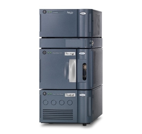 Waters ACQUITY UPLC (Fig from Waters)
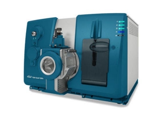 AB Sciex Triple Quad 6500+ (Fig from Sciex)
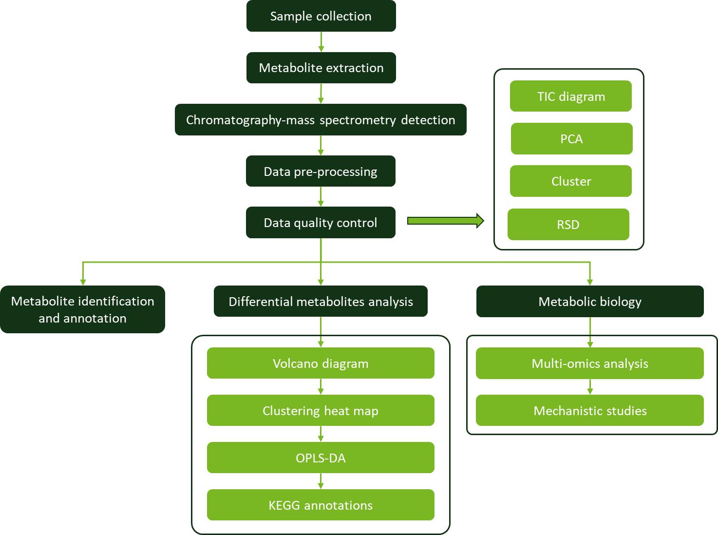
Workflow for Xanthophylls Analysis Service
Sample Requirements for Xanthophylls (Contain Oxygen) Assay
| Sample Type |
Minimum Amount Required |
Storage Conditions |
Notes |
| Plant Leaves |
≥100 mg (fresh) / ≥50 mg (dried) |
Fresh: -80°C, Dried: Room temp |
Avoid prolonged exposure to light and oxygen. |
| Algae Cultures |
≥10 mL |
-80°C |
Pellet cells by centrifugation if possible. |
| Fruits & Vegetables |
≥50 g |
-20°C |
Store whole or homogenized. |
| Food Products (Eggs, Dairy, Oils, etc.) |
≥50 g / ≥10 mL (liquid) |
-20°C |
Store in airtight containers. |
| Serum / Plasma |
≥200 µL |
-80°C |
Use EDTA or heparin tubes for collection. |
| Fermented Products |
≥10 mL |
-20°C |
Store in sterile, sealed containers. |
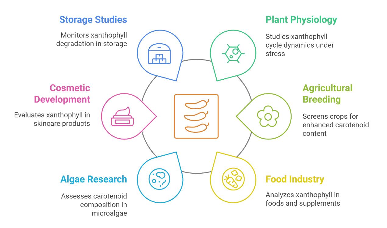
Applications of Xanthophylls Analysis
Case. Red light-transmittance bagging promotes carotenoid accumulation through xanthophylls esterification during the ripening of blood orange fruit
Background:
The importance of carotenoids and light was investigated.
This study aimed to explore the effects of different light - transmittance bagging on carotenoid metabolism in 'Moro' blood orange during ripening.
Samples:
Healthy and uniform 'Moro' blood oranges (C. sinensisL. Osbeck) from five plants.
Technical methods procedure:
Carotenoid extraction and analysis was resuspended and analyzed by UPLC - APCI - MS/MS.
RNA extraction and RT - qPCR: Total RNA was extracted from the frozen pulp, and first - strand cDNA was synthesized. RT - qPCR was performed to analyze the expression levels of candidate genes.
Homologous gene screening and phylogenetic analysis: PF00657 was used to screen homologous genes of GDSL Esterase/Lipase in citrus, and a phylogenetic analysis was conducted.
Pairwise comparisons of esterified xanthophylls levels were done, and partial Mantel correlations between esterified xanthophylls and candidate gene expression levels were computed.
Results:
50 carotenoids were identified in 'Moro' blood orange pulp, including 13 free forms and 37 esterified xanthophylls. Violaxanthin derivatives were the most common.
Red LTB promoted carotenoid accumulation, especially xanthophylls.
Blue LTB negatively regulated carotenoid accumulation via carotene metabolism.
Red LTB promoted xanthophylls esterification. The levels and proportions of esterified lutein, β - cryptoxanthin, and violaxanthin were significantly higher in red LTB.
CsAt1g54570 was significantly correlated with the level of esterified β - cryptoxanthins.
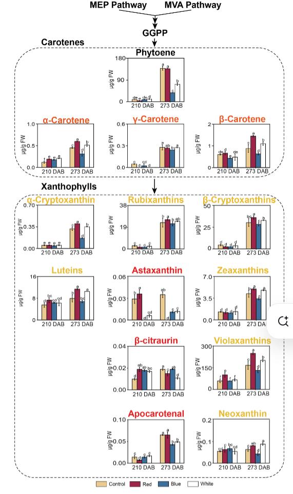 Changes in identified carotenes and xanthophylls content during the ripening of blood orange under bagging with different light transmittance.
Reference
- Yang C, et al. "Red light-transmittance bagging promotes carotenoid accumulation through xanthophylls esterification during the ripening of blood orange fruit." Food Chemistry (2023) 404: 134578. https://doi.org/10.1016/j.foodchem.2022.134578

