
Metabolic flux analysis (MFA) has become a powerful tool to provide important quantitative information about the metabolism of biological systems that cannot be obtained from other "omics" methods. The development of analytical techniques such as mass spectrometry (MS) and nuclear magnetic resonance (NMR) spectroscopy and the use of dedicated software tools has greatly increased the precision and accuracy of flux analysis. To date, this method is widely established and applied in microorganisms. Mammalian cells are key platforms for biopharmaceutical production and biomedical research and their metabolism is complex.
Service offering at Creative Proteomics
MFA quantitatively characterizes cellular fluxes to understand metabolic phenotypes and functional behavior in response to environmental and/or genetic perturbations. Isotope labeling experiments help MFA by providing information on intracellular fluxes, especially through parallel and circular pathways. Based on the optimized analytical methods, software packages, and improved computational modeling, Creative Proteomics is committed to providing MFA services aimed at quantifying the fluxes of complex eukaryotic systems. We are proud to offer customized and one-stop MFA services in mammalian cells, including experimental design, labeling experiment, sample preparation, NMR-based or MS-based metabolic flux analysis, and data analysis.
Specifically, we focus on the following lists, but are not limited to:
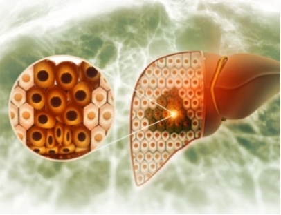
The liver is the primary organ responsible for metabolizing drugs and toxins in the body. Identification of hepatocyte metabolic pathways will help to monitor changes under physiological and pathophysiological perturbations. In addition, primary hepatocytes isolated from the liver are useful tools for assessing metabolism, drug interactions, hepatotoxicity and transporter activity in vitro. MFA provides a comprehensive view of the intracellular distribution of metabolic fluxes and can be applied to hepatocyte metabolism studies.
Although the brain represents only 2% of the total body weight, it accounts for 20% of an individual's resting energy expenditure. In addition, metabolic alterations have been shown to strongly influence the initiation and progression of many neurodegenerative diseases. The rate of glucose and oxygen metabolism in brain cells is declining during the normal aging process and is further exacerbated in the neurodegenerative diseases described above. In addition to advancing our understanding of how the brain functions and adapts to environmental demands, MFA may also further elucidate the mechanisms by which age-related neurodegenerative diseases occur in the human brain.


Cancer metabolism is significantly altered from normal cellular metabolism. Reprogrammed cellular metabolism allows cancer cells to maintain high proliferation rates and adapt to changing microenvironments. Over the past decade, stable-isotope tracing and network analysis have become powerful tools to reveal differentially activated metabolic pathways in cancer cells. With rapid advances in cancer research, it is becoming increasingly clear how cancer cells rewire their metabolism to promote the proliferation of more cancer cells.
Applications of our service
- Analysis and optimization of cell culture processes, involving cultivation conditions, substrates, or other factors affecting cellular metabolism
- Facilitate the characterization of drug metabolism in mammalian cells
- Identify cancer biomarkers
- Elucidate complex mechanisms of various cancers
- Contribute to precision treatment with pharmacometabolomics