Get Your Custom Quote

Online Inquiry

Strengthen your biomarker discovery pipeline with integrated proteomics and multi-omics services. Our approach enhances specificity, reliability, and translational relevance.

Get Your Custom Quote

Biomarker Identification Analysis

Uncover reliable and biologically meaningful biomarkers from complex samples with confidence. Our biomarker identification services combine mass spectrometry-based proteomics and integrated multi-omics analysis to address key challenges such as low specificity, poor reproducibility, and limited translational relevance. Backed by rigorous experimental workflows and expert data interpretation, we deliver robust biomarker candidates that support high-quality life science and translational research.

  • High-confidence discovery: Quantitative MS-based proteomics integrated with multi-omics to improve biomarker specificity and robustness
  • End-to-end workflow: From discovery to targeted validation using PRM, immunoassays, and custom antibody-based strategies
  • Proven expertise: Developed by experienced scientists following established proteomics and multi-omics research standards
Creative Proteomics' biomarker identification service.

Why Biomarker Identification is Important?

Biomarkers are measurable biological indicators that reflect physiological states, pathological processes, or responses to interventions. They provide essential insights into molecular events and disease mechanisms. Accurate biomarker identification is pivotal for life science research and translational studies. Biomarkers can span nucleic acids, proteins, metabolites, epigenetic modifications, and cellular components.

Reliable biomarkers are characterized by specificity and reproducibility. They often correlate with disease progression or distinct biological phenotypes. In the context of precision research, biomarkers guide the selection of targets for molecular studies, stratify biological samples, and enhance understanding of complex molecular networks.

Creative Proteomics' biomarker discovery workflow.

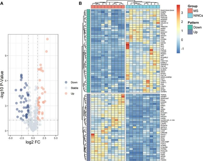
Figure 1. The 14 cancer hallmarks-based biomarkers (Zhou Y, et al., 2024).

MS-Based Proteomics for Biomarker Identification

High-Resolution Protein Profiling

Mass spectrometry enables researchers to simultaneously measure the abundance, structure, and modifications of thousands of proteins. High-resolution instruments generate precise and reproducible protein data. These tools can detect even proteins present at very low levels, which are often the most important biomarkers.

Quantitative Accuracy

Quantitative proteomics strategies enable the accurate comparison of protein levels across different conditions or patient groups. Methods like label-free quantification or stable isotope labeling provide clear numerical values for protein abundance.

Detection of PTMs

Proteins often undergo chemical changes after they are produced, such as phosphorylation or glycosylation. Mass spectrometry can detect these modifications, revealing functional changes that may indicate disease processes.

Quantitative Proteomics Strategies for Biomarker Discovery

Quantitative proteomics provides the foundation for identifying reliable biomarkers by accurately measuring protein abundance across biological samples.

Integrating Multi-Omics to Improve Biomarker Specificity

Multi-omics integration combines complementary layers of molecular information, thereby improving the specificity and robustness of biomarker discovery. Key multi-omics modalities include

Creative Proteomics provides integrated analysis:

Integrative Analysis of PTMs and Metabolomics

Integrative Analysis Services of Proteomics and Phosphoproteome

Integrative Analysis Services of Proteomics-Acetyl

Validation Strategies for Biomarkers

Validation ensures that the biomarkers are robust indicators of the intended biological state and suitable for downstream applications.

Our Biomarker Discovery Workflow

Workflow for biomarker discovery.

Data Deliverables & What You Receive

Applications Across Life Science and Translational Research

Sample Requirements

Sample Type Recommended Format Notes
Tissue Fresh, frozen, or FFPE Minimum quantity depends on tissue type.
Blood/Plasma/Serum EDTA, heparin, or citrate collection Ensure proper storage to preserve protein integrity.
Cell Lines Whole cells or lysates Harvest at exponential growth phase.
Biofluids Urine, cerebrospinal fluid, saliva Pre-treatment may be required.
Experimental Models Homogenates, organoids, xenografts Standardized collection for reproducibility.

Why Choose Creative Proteomics

FAQ

Q1: Why do some biomarker candidates fail validation or translation?

A1: Many biomarker candidates discovered in early studies do not hold up in later work. Common reasons include small or unbalanced sample sets, inconsistent data across experiments, and the absence of independent confirmation. To overcome these limitations, robust statistical analysis, testing in separate sample groups, and validation using complementary methods are essential.

Q2: Can multi-omics analysis identify low-abundance biomarkers?

A2: Yes. Combining data from genomics, transcriptomics, proteomics, and metabolomics enables the detection of small but biologically significant changes, even when the molecules of interest are present at very low levels.

Q3: Can biomarker discovery identify dynamic or temporal changes in disease progression?

A3: Yes. Collecting samples over time and analyzing them using quantitative proteomics and integrated multi-omics enables the tracking of molecular changes as they occur. This approach helps identify dynamic biomarkers that reflect disease progression or biological responses to experimental interventions.

Demo

Demo: LC‑MS/MS Based Metabolomics and Proteomics Reveal Candidate Biomarkers and Molecular Mechanism of Early IgA Nephropathy

This integrative study combined untargeted LC-MS/MS proteomics and metabolomics to investigate plasma samples from patients with IgA nephropathy versus those from healthy controls. Differentially expressed proteins and metabolites were identified and analyzed with machine learning. Key candidates such as PRKAR2A, IL6ST, and SOS1 were highlighted. Independent validation showed strong classification performance, underscoring the value of combined omics for early biomarker discovery.

Results of Proteomic analysis for IgAN.

Figure 2. Proteomic analysis of IgAN (Zhang D, et al., 2022).

Results for DEPs GO and KEGG pathway analysis.

Figure 3. GO terms and KEGG pathway analysis for DEPs (Zhang D, et al., 2022).

Case Study

Case: Discovery of Novel Biomarkers for Diagnosing and Predicting the Progression of Multiple Sclerosis Using TMT‑Based Quantitative Proteomics

Abstract

Multiple sclerosis (MS) is a chronic inflammatory and neurodegenerative condition of the central nervous system. Biomarker discovery for MS is challenging due to disease complexity and overlapping clinical features with other neurological disorders. Reliable protein biomarkers in cerebrospinal fluid (CSF) and plasma can improve molecular insights and support stratification of disease states.

The study aimed to identify protein biomarkers that differentiate MS patients from non‑inflammatory neurological controls using quantitative proteomics. The authors also sought to evaluate whether candidate proteins could distinguish disease progression phenotypes.

Methods

  • Employed an isobaric mass tag labeling strategy (Tandem Mass Tag, TMT) coupled with high‑resolution mass spectrometry to quantify protein expression.
  • Differentially expressed proteins (DEPs) were identified through proteomic bioinformatics.
  • Candidate proteins were further validated in a larger cohort of 160 samples (paired CSF and plasma) using enzyme‑linked immunoassay (ELISA).
  • Receiver operating characteristic (ROC) curves assessed diagnostic relevance.

Results

  • Out of 343 quantified proteins, 83 were differentially expressed between MS patients and controls.
  • Functional enrichment analysis revealed involvement in processes such as platelet degranulation and signaling pathways relevant to immune regulation.
  • Three proteins emerged as central in the protein–protein interaction network.
  • ELISA validation confirmed significant upregulation of IGFBP7 and downregulation of SST in CSF from MS patients.
  • IGFBP7 also demonstrated potential to discriminate between relapsing‑remitting and secondary progressive MS phenotypes.
Results for GO and KEGG pathway analyses.

Figure 4. GO and KEGG pathway analyses of MS-related proteins.

Results for volcano plot and heatmap.

Figure 5. Volcano plot and heatmap of patients with MS vs NINCs.

Results for the DEPs interaction network.

Figure 6. Interaction network of the DEPs.

Conclusion

High-throughput quantitative proteomics, coupled with TMT labeling, can reveal robust protein biomarkers in complex clinical samples. IGFBP7 and SST show promise as protein indicators associated with MS pathology, with IGFBP7 potentially serving as a key marker for disease classification and progression.

Related Services

References

For research purposes only, not intended for clinical diagnosis, treatment, or individual health assessments.

Tell Us About Your Project