Antibody-Based Immunoassay Detection of DNA and RNA Modifications for Accessible, High-Throughput Epigenetic and Epitranscriptomic Analysis
Chemical modifications of DNA and RNA — including DNA methylation (5mC, 5hmC, 5fC, 5caC), RNA methylations (m⁶A, m⁵C, m¹A, m⁷G), pseudouridine, and oxidative damage products (8-oxo-dG) — are fundamental regulators of gene expression, genome stability, and cellular function, with established roles in development, differentiation, and disease pathogenesis including cancer, neurological disorders, cardiovascular disease, and metabolic conditions. Quantifying these modifications is essential for understanding their biological functions, establishing their utility as disease biomarkers, and evaluating the effects of therapeutic interventions on the epigenome and epitranscriptome. While LC-MS/MS provides the gold standard for absolute quantification and sequencing-based methods offer transcriptome-wide site resolution, immunoassay-based detection using modification-specific antibodies offers distinct advantages for specific research contexts: lower cost per sample, higher throughput (96-well plate format), minimal sample preparation requirements, accessibility without specialized mass spectrometry expertise, and compatibility with standard laboratory equipment (plate readers, chemiluminescent imagers, qPCR instruments).
Four complementary immunoassay formats are available to address different research needs. Competitive ELISA provides quantitative colorimetric detection in a high-throughput 96-well format with standard absorbance readout, ideal for large cohort studies where sample numbers exceed 50–100. Dot blot offers the simplest and most cost-effective format for rapid modification screening with chemiluminescent detection, requiring only a membrane, antibodies, and an imaging system. Fluorometric assays provide enhanced sensitivity through fluorescent signal amplification, particularly valuable for low-abundance modifications or limited sample amounts. qPCR-compatible detection — combining modification-specific antibody enrichment with quantitative PCR readout — enables locus-specific quantification of modifications at defined genomic (MeDIP-qPCR, hMeDIP-qPCR) or transcriptomic (m⁶A-qPCR, m⁵C-qPCR) regions, bridging the gap between global quantification and sequencing-based site mapping. For comprehensive modification analysis at the nucleoside level, our m⁶A Modification LC-MS Analysis and Oxidative DNA/RNA Damage Assay services provide orthogonal LC-MS/MS quantification for cross-platform validation and deeper mechanistic characterization.
Find Your Solution: Research Goal → DNA/RNA Modification Immunoassay Strategy
| Your Research Goal |
Recommended Approach |
Key Techniques |
| High-throughput global quantification of DNA methylation (5mC, 5hmC) or RNA modification (m⁶A, m⁵C) levels across large clinical cohorts (50–500+ samples) |
Competitive colorimetric ELISA in 96-well plate format |
DNA/RNA extraction, denaturation, plate coating with modified nucleoside conjugate or antibody, competitive binding with sample modification, HRP/TMB colorimetric detection, standard curve-based quantification at 450 nm absorbance readout |
| Rapid, low-cost screening of RNA modification levels (m⁶A, m⁵C, Ψ, m¹A) in total RNA or mRNA across multiple samples or time points |
Dot blot with chemiluminescent detection |
RNA denaturation and direct spotting on nitrocellulose membrane, UV crosslinking, modification-specific primary antibody incubation, HRP-conjugated secondary antibody, ECL chemiluminescent imaging, densitometric quantification normalized to total RNA loading (methylene blue staining) |
| Sensitive detection of low-abundance DNA/RNA damage modifications (8-oxo-dG, 8-oxo-dA, etheno-dA) in biofluid or tissue lysate samples |
Fluorometric competitive ELISA with enhanced sensitivity |
Sample DNA/RNA extraction and enzymatic digestion, competitive binding format with fluorescent detection (Alexa Fluor/HRP-fluorophore), plate reader quantification with excitation/emission at optimized wavelengths, lower limits of detection compared to colorimetric ELISA |
| Locus-specific quantification of 5mC or 5hmC at defined gene promoters or genomic regions |
Antibody enrichment coupled with qPCR (MeDIP-qPCR, hMeDIP-qPCR) |
Genomic DNA fragmentation (sonication or enzymatic), 5mC/5hmC-specific antibody immunoprecipitation, qPCR with primers targeting regions of interest, percent input normalization, enrichment fold-change calculation relative to IgG control or input DNA |
| Locus-specific quantification of m⁶A or m⁵C at defined transcript regions or specific gene mRNAs |
m⁶A-qPCR or m⁵C-qPCR with antibody enrichment |
mRNA purification (polyA selection), fragmentation, m⁶A/m⁵C-specific antibody immunoprecipitation, qPCR with primers targeting specific transcript regions, normalization to input or unmodified control regions, enrichment fold-change calculation |
| Multi-modality modification profiling: combining global quantification with locus-specific analysis and orthogonal LC-MS/MS validation |
Integrated workflow: ELISA or dot blot screening → qPCR locus-specific follow-up → LC-MS/MS validation |
Tiered approach: global screening by ELISA/dot blot identifies modification-level changes, qPCR-based locus-specific analysis identifies modified genes/transcripts, LC-MS/MS provides absolute quantification and cross-platform validation of key findings |
Complementary Immunoassay Platforms for DNA and RNA Modification Detection
Our DNA/RNA modification immunoassay service portfolio encompasses four complementary detection platforms, each optimized for specific throughput, sensitivity, cost, and locus-specific resolution requirements. Platform selection depends on the research question, modification target, sample type, sample throughput, and desired quantitative resolution.
Competitive ELISA for Quantitative High-Throughput Global Modification Detection
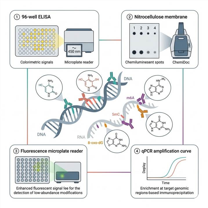
Competitive ELISA is our primary platform for high-throughput quantitative detection of global DNA and RNA modification levels in a 96-well plate format. In this format, the sample DNA or RNA is denatured and immobilized on the plate surface, or alternatively, a modified nucleoside conjugate is coated on the plate and competes with sample modification for antibody binding. A modification-specific primary antibody (validated for ELISA) is added, followed by an HRP-conjugated secondary antibody and TMB colorimetric substrate, with absorbance measured at 450 nm. Quantification is achieved by reference to a standard curve constructed from known concentrations of the target modified nucleoside. The competitive ELISA format provides quantitative results for modification levels expressed as a percentage of total nucleosides (e.g., %5mC, %5hmC, %m⁶A) with typical limits of detection of 0.01–0.1% modification fraction, depending on the target. Throughput of 80–200 samples per week (including standards and controls in each plate) makes ELISA the method of choice for large cohort studies in clinical epigenetics and epitranscriptomics research.
Dot Blot for Rapid, Low-Cost Modification Screening with Minimal Sample Preparation
Dot blot provides the simplest and most cost-effective format for rapid screening of DNA and RNA modification levels. In this format, denatured DNA or RNA samples are directly spotted onto a nitrocellulose or PVDF membrane, UV-crosslinked or baked to immobilize the nucleic acids, incubated with modification-specific primary antibody, detected with HRP-conjugated secondary antibody and ECL chemiluminescent substrate, and imaged on a chemiluminescence imaging system (ChemiDoc, Amersham Imager, or equivalent). Total nucleic acid loading is normalized by methylene blue or SYBR Gold staining of the same membrane, providing semi-quantitative modification levels expressed as chemiluminescent signal per unit nucleic acid. Dot blot is particularly valuable for pilot experiments, time-course studies comparing modification dynamics across multiple time points, and studies with limited sample numbers where the investment of a full ELISA plate is not justified. The minimal sample preparation (DNA/RNA extraction, denaturation, and spotting) makes dot blot accessible for laboratories without specialized equipment beyond a standard chemiluminescence imager.
Fluorometric Competitive ELISA for Enhanced-Sensitivity Detection of Low-Abundance Modifications
For modifications present at very low levels in biological samples — including 8-oxo-dG (typically 0.1–10 lesions per 10⁶ nucleotides in normal cells), 5fC and 5caC (intermediates in active DNA demethylation at 0.001–0.01% of total cytosines), and etheno-DNA adducts (lipid peroxidation damage products at sub-fmol levels) — our fluorometric competitive ELISA platform provides enhanced sensitivity through fluorescent signal detection. This format replaces colorimetric HRP/TMB detection with fluorophore-conjugated secondary antibodies (Alexa Fluor 488, 555, or 647) or HRP-fluorophore substrate systems that produce fluorescent rather than chromogenic signals, achieving 3–10-fold improvements in detection limits compared to standard colorimetric ELISA. Quantification is performed using a fluorescence microplate reader with appropriate excitation and emission filters, and results are calculated by reference to a standard curve as in the colorimetric format.
Antibody Enrichment with qPCR Readout for Locus-Specific Modification Quantification
For researchers who need to move beyond global modification levels and quantify modifications at specific genomic or transcriptomic regions — such as 5mC or 5hmC at a specific gene promoter, or m⁶A at a specific mRNA transcript — our antibody enrichment coupled with quantitative PCR (qPCR) platform provides locus-specific modification quantification. In this workflow, DNA (for 5mC, 5hmC) or RNA (for m⁶A, m⁵C) is extracted, fragmented to 200–500 bp fragments, incubated with modification-specific antibody to immunoprecipitate the modified fragments, and the enriched DNA/RNA is analyzed by qPCR using primers targeting the genomic or transcriptomic regions of interest. Enrichment is quantified as percent input or fold-change relative to a negative control (IgG or unmodified control region). This approach bridges the gap between global ELISA/dot blot quantification and sequencing-based genome-wide mapping, providing quantitative modification data at specific loci relevant to the researcher's hypotheses without the cost and data complexity of genome-wide sequencing. For dedicated genome-wide modification profiling, complementary sequencing services are available through our partner platforms.
Why Choose Our DNA/RNA Modification Immunoassay Services
Four Complementary Immunoassay Platforms Covering All Detection Needs from Global Screening to Locus-Specific Quantification
We offer the most comprehensive immunoassay service portfolio for DNA and RNA modification detection, spanning competitive ELISA (colorimetric, high-throughput), dot blot (rapid, low-cost screening), fluorometric ELISA (enhanced sensitivity for low-abundance modifications), and antibody enrichment with qPCR readout (locus-specific quantification). This breadth ensures that we can match the optimal detection platform to each specific research question — from global modification screening across large clinical cohorts to targeted quantification at specific genes or transcripts — within a single service relationship, eliminating the need to coordinate multiple providers for different assay formats.
Validated Modification-Specific Antibody Portfolio with Rigorous Quality Control
Our modification-specific antibody panel includes well-characterized monoclonal and polyclonal antibodies against 15+ DNA and RNA modifications — including 5mC (clone 33D3), 5hmC (clone RM236), 5fC, 5caC, 6mA, N7-meG, O⁶-meG, 8-oxo-dG (clone 483.15), 8-oxo-dA, m⁶A (clones 17-3-1-1, 202003), m⁵C (clone 4G3), pseudouridine (clone 20-100-1), m¹A, m⁷G, ac⁴C, inosine, and 8-oxo-rG — with each antibody validated for its specific immunoassay format (ELISA, dot blot, immunoprecipitation) through specificity testing against unmodified nucleosides, modified nucleoside competitors, and biological positive/negative control samples. Lot-to-lot consistency and cross-reactivity profiles are documented for each antibody.
Comprehensive DNA and RNA Modification Coverage from a Single Service Provider
Our service covers the full spectrum of biologically important DNA and RNA modifications — epigenetic DNA modifications (5mC, 5hmC, 5fC, 5caC, 6mA), DNA damage and adduct modifications (8-oxo-dG, 8-oxo-dA, N7-meG, O⁶-meG, etheno-dA), epitranscriptomic RNA modifications (m⁶A, m⁵C, pseudouridine, m¹A, m⁷G, ac⁴C, inosine, 2'-O-methylations), and RNA damage (8-oxo-rG) — from the same DNA/RNA samples, enabling integrated analysis of multiple modification types within a single study. This comprehensive coverage is particularly valuable for multi-omics studies investigating coordinated changes across the epigenome and epitranscriptome, or for studies of DNA damage and repair where multiple damage products need to be quantified simultaneously.
Integrated Multi-Modality Workflow with Optional LC-MS/MS Cross-Platform Validation
We offer a unique integrated workflow that connects immunoassay-based screening with LC-MS/MS-based absolute quantification for cross-platform validation of key findings. Researchers can begin with high-throughput ELISA or dot blot screening to identify modification-level changes across all samples, then select the most informative samples or modifications for orthogonal validation by LC-MS/MS — combining the throughput and accessibility of immunoassays with the gold-standard quantitative accuracy of isotope dilution mass spectrometry. This tiered approach optimizes the balance between throughput and quantitative rigor, ensuring that published findings are supported by complementary analytical methods.
Workflow: From Sample to Quantified DNA/RNA Modification Data
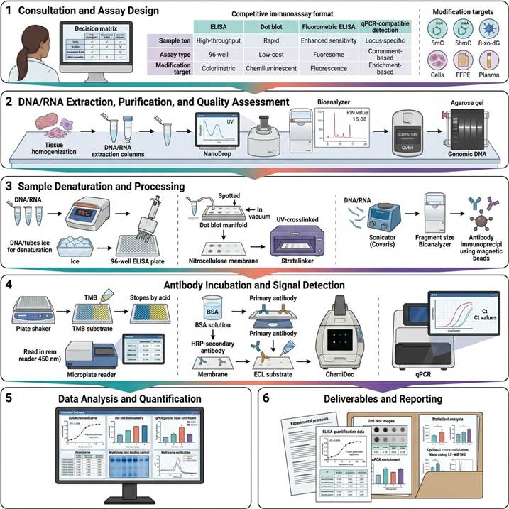
Step 1: Consultation and Assay Design
We consult with you to define the DNA/RNA modification immunoassay strategy based on modification targets (5mC, 5hmC, m⁶A, 8-oxo-dG, custom panel), sample type (gDNA from tissue/cells/FFPE, total RNA, mRNA, cfDNA, biofluid), throughput requirements (10–500+ samples), detection format (ELISA, dot blot, fluorometric, qPCR-compatible), and whether locus-specific quantification is required. We provide a detailed project proposal with platform recommendations, expected sensitivity, and sample requirements.
Step 2: DNA/RNA Extraction, Purification, and Quality Assessment
DNA or RNA is extracted from provided samples using optimized protocols appropriate for the sample type (tissue, cells, FFPE, plasma, serum). DNA/RNA quantity is assessed by NanoDrop and Qubit fluorometry, purity by A260/A280 and A260/A230 ratios, and integrity by agarose gel electrophoresis or Bioanalyzer (RIN value for RNA). For genomic DNA, RNase treatment is performed to eliminate RNA contamination. For RNA, DNase treatment removes genomic DNA contamination. Samples are normalized to uniform concentration before assay.
Step 3: Sample Denaturation and Processing
Depending on the assay format: for ELISA, DNA/RNA is denatured by heating (95–100°C, 5–10 minutes) followed by rapid cooling on ice to maintain single-stranded conformation, then loaded onto the ELISA plate at the optimized concentration. For dot blot, samples are denatured, serially diluted if needed, and spotted onto nitrocellulose membrane followed by UV crosslinking. For qPCR-compatible detection, DNA/RNA is fragmented by sonication or enzymatic digestion to 200–500 bp fragments for efficient antibody immunoprecipitation.
Step 4: Antibody Incubation and Signal Detection
For ELISA: plate is blocked (BSA or milk), incubated with modification-specific primary antibody (1 hour, room temperature or overnight at 4°C), washed, incubated with HRP-conjugated secondary antibody (1 hour), washed, and developed with TMB substrate. Absorbance is measured at 450 nm. For dot blot: membrane is blocked, incubated with primary antibody (overnight, 4°C), washed, incubated with HRP-secondary, washed, and developed with ECL substrate for chemiluminescent imaging. For fluorometric assays: fluorophore-conjugated detection replaces HRP/TMB with appropriate excitation/emission wavelength readout.
Step 5: Data Analysis and Quantification
For ELISA: absorbance values are converted to modification percentage using the standard curve (four-parameter logistic regression). For dot blot: chemiluminescent signal intensity is quantified by densitometry and normalized to total nucleic acid loading (methylene blue staining intensity). For qPCR-compatible detection: enrichment is calculated as percent input using the ΔΔCt method relative to input DNA/RNA or as fold-change relative to IgG control. Statistical analysis includes appropriate tests for group comparisons with multiple testing correction where applicable.
Step 6: Deliverables and Reporting
Comprehensive immunoassay report including: complete experimental protocols with DNA/RNA preparation, assay conditions, and antibody specifications, quantitative modification data (% modification or signal intensity per unit nucleic acid) for all samples with calibration curves and quality control metrics, statistical analysis results with group comparisons and data visualization (bar charts, box plots, scatter plots), and optional cross-platform validation data when integrated with LC-MS/MS analysis. All raw data (plate reader absorbance values, chemiluminescent images, qPCR amplification curves) are provided in standard formats.
Applications in DNA/RNA Modification Immunoassay Research
Antibody-based immunoassay detection of DNA and RNA modifications has become a widely adopted approach for accessible, high-throughput epigenetic and epitranscriptomic analysis across diverse research fields. The following application areas represent the most active and impactful use cases for our immunoassay platforms.
Clinical Epigenetics: Global DNA Methylation and Hydroxymethylation Quantification in Disease Cohorts
Global DNA methylation (5mC) and hydroxymethylation (5hmC) levels are emerging as diagnostic, prognostic, and predictive biomarkers in cancer, neurological disorders, and cardiovascular disease. Competitive ELISA quantification of 5mC and 5hmC in genomic DNA from clinical tissue samples, blood, or cell-free DNA enables rapid assessment of global epigenetic changes across large patient cohorts. The 96-well plate format provides the throughput needed for studies involving 50–500+ clinical samples, with the sensitivity to detect modification changes as subtle as 1–2% absolute difference in 5hmC content between disease and control groups. For example, global 5hmC reduction is a well-established epigenetic hallmark of many cancer types — including colorectal, breast, prostate, lung, and liver cancer — and ELISA-based 5hmC quantification from FFPE tissue DNA or circulating cell-free DNA has been validated as a clinically accessible biomarker approach in multiple independent studies.
Epitranscriptomics: Global RNA Modification Screening Across Biological Conditions and Drug Treatments
Dot blot and ELISA quantification of m⁶A, m⁵C, pseudouridine, and m¹A in total RNA or mRNA provides a rapid, cost-effective entry point for epitranscriptomics research. Before investing in transcriptome-wide m⁶A sequencing or comprehensive LC-MS/MS analysis, researchers can use dot blot screening to determine whether modification-level changes occur under their experimental conditions, across which time points the most significant changes are observed, and which RNA modifications show the most robust responses to treatment. This screening approach is particularly valuable for pilot studies, time-course experiments, and multi-condition drug treatment studies where the experimental space is too large for immediate sequencing or LC-MS/MS investment. For deeper quantification, our RNA Modification Quantification by LC-MS/MS service provides orthogonal absolute quantification for key findings identified in immunoassay screens.
DNA Damage and Oxidative Stress Biomarker Quantification in Toxicology and Disease Research
8-Oxo-7,8-dihydro-2'-deoxyguanosine (8-oxo-dG) is the most widely studied biomarker of oxidative DNA damage, with established associations with aging, cancer, neurodegeneration, cardiovascular disease, diabetes, and environmental toxin exposure. Competitive ELISA quantification of 8-oxo-dG in DNA hydrolysates from tissue, blood, or urine provides a sensitive, high-throughput readout of oxidative stress levels that is accessible without specialized LC-MS/MS instrumentation. Our fluorometric competitive ELISA platform provides enhanced sensitivity for 8-oxo-dG quantification from limited sample amounts (as low as 0.5–5 μg DNA per well) and is compatible with both DNA and RNA samples (8-oxo-rG quantification). For orthogonal validation, our Oxidative DNA/RNA Damage Assay provides LC-MS/MS-based absolute quantification of multiple oxidative damage products in a single analytical run.
Drug Development: Epigenetic Drug Target Engagement and Pharmacodynamic Biomarker Assessment
Epigenetic drugs — including DNA methyltransferase inhibitors (DNMTi, e.g., decitabine, azacitidine), histone deacetylase inhibitors (HDACi), and TET activators — modulate DNA modification levels as their primary mechanism of action. ELISA-based quantification of global 5mC and 5hmC levels in treated vs. control cells or tissues provides a direct pharmacodynamic readout of drug target engagement: DNMT inhibitors cause global 5mC reduction, while TET activators increase 5hmC levels. Similarly, drugs targeting RNA modification enzymes (METTL3 inhibitors, FTO inhibitors) can be evaluated for their effects on global m⁶A levels using m⁶A ELISA or dot blot. The high throughput and low cost of immunoassay-based detection make it well-suited for dose-response studies, time-course experiments, and combination therapy screening where multiple conditions need to be evaluated in parallel. For deeper mechanistic characterization of target engagement, our Reactive Cysteine Target Engagement Assay provides complementary chemoproteomic target engagement analysis.
Locus-Specific Modification Quantification: Targeted Epigenetic and Epitranscriptomic Validation
Following identification of global modification changes by ELISA or dot blot, or following genome-wide modification mapping by sequencing, researchers often need to validate modification levels at specific genes or transcripts of interest. Our antibody enrichment coupled with qPCR (MeDIP-qPCR, hMeDIP-qPCR, m⁶A-qPCR) provides targeted locus-specific modification quantification without the cost and complexity of genome-wide sequencing. This approach enables: validation of differentially methylated promoters identified in 5mC sequencing studies; quantification of 5hmC changes at specific gene bodies in cancer versus normal tissue; assessment of m⁶A enrichment at specific oncogene or tumor suppressor transcripts; and targeted analysis of modification changes at developmentally regulated or disease-associated genomic loci across multiple biological replicates and time points. For comprehensive genome-wide modification profiling, complementary sequencing services are available through our partner platforms.
Case Study: ELISA-Based Quantification of 5-Hydroxymethylcytosine in Colorectal Cancer FFPE Tissues Reveals AMPK/TET2/5-hmC Axis in Obesity-Related Carcinogenesis
In a 2023 study published in Scientific Reports (CC BY 4.0), Kon et al. employed competitive ELISA-based 5-hydroxymethylcytosine (5hmC) quantification from FFPE tissue-derived genomic DNA to investigate the relationship between metabolic alterations, TET enzyme expression, and global DNA hydroxymethylation in obesity-related colorectal cancer — demonstrating the utility of immunoassay-based DNA modification detection in clinical epigenetics research.
Background: Obesity is an established risk factor for colorectal cancer (CRC), yet the molecular mechanisms linking metabolic dysregulation to epigenetic changes that drive CRC development were incompletely understood. Global loss of 5hmC — an epigenetic mark generated by TET enzyme-mediated oxidation of 5mC — has been identified as a hallmark of many cancer types, but its relationship with metabolic alterations in obesity-related CRC had not been systematically characterized. The authors hypothesized that obesity-induced metabolic reprogramming suppresses TET enzyme expression and activity, leading to reduced 5hmC levels that contribute to CRC development through epigenetic dysregulation of tumor suppressor and oncogene pathways.
Approach: Genomic DNA was extracted from formalin-fixed, paraffin-embedded (FFPE) colorectal cancer tissues and adjacent non-cancerous tissues from 40 CRC patients stratified by obesity status (obese vs. non-obese). Global 5hmC levels in genomic DNA were quantified using a competitive 5hmC ELISA kit, with results expressed as percentage of 5hmC relative to total cytosine content. TET2 mRNA expression was analyzed by RT-qPCR in matched tissue samples, and TET2 protein expression was assessed by immunohistochemistry (IHC). Functional experiments in CRC cell lines (HCT116, HT-29, DLD-1) examined the effects of glucose and insulin treatment on TET2 expression and 5hmC levels, and the AMPK activator AICAR was used to evaluate the role of AMPK signaling in TET2 regulation.
Key Findings:
- Reduced 5hmC in CRC tissues: Competitive ELISA quantification revealed that global 5hmC levels were significantly decreased in colorectal cancer tissues compared to adjacent non-cancerous tissues (p < 0.01), consistent with the established observation that 5hmC loss is a widespread epigenetic feature of malignant transformation, and validating the ELISA platform for detecting clinically relevant epigenetic changes in FFPE-derived DNA
- Obesity-associated 5hmC reduction: In obese CRC patients, 5hmC levels in tumor tissues were significantly lower than in non-obese CRC patients (p = 0.03), while no significant difference in 5hmC was observed between obese and non-obese groups in adjacent normal tissues (p = 0.46), suggesting that obesity specifically exacerbates cancer-associated epigenetic dysregulation
- TET2 suppression as the mechanistic link: TET2 mRNA and protein expression were significantly reduced in CRC tissues compared to normal tissues, with further reduction in obese patients. In CRC cell lines, glucose and insulin treatment — modeling the metabolic environment of obesity — significantly suppressed TET2 expression and reduced 5hmC levels, while AMPK activation by AICAR restored TET2 expression and 5hmC levels, establishing the AMPK/TET2/5-hmC signaling axis as a mechanistic link between metabolic alterations and epigenetic dysregulation in obesity-related CRC
- ELISA as a clinical epigenetics tool: The competitive 5hmC ELISA platform provided quantitative, reproducible 5hmC measurements from FFPE-derived genomic DNA — the most commonly available clinical tissue archive format — demonstrating that immunoassay-based DNA modification detection is directly compatible with clinical pathology workflows and can be applied to retrospective analysis of archived tissue samples with known clinical outcomes
- 5hmC as a pharmacodynamic biomarker: The finding that AMPK activation by AICAR restored TET2 expression and 5hmC levels in CRC cells suggests that 5hmC ELISA could serve as a pharmacodynamic biomarker for evaluating AMPK-targeted therapeutic interventions in clinical trials, with the ELISA format providing the throughput and accessibility needed for clinical sample analysis
Significance: This study established that obesity-related metabolic alterations suppress the AMPK/TET2/5-hmC axis in colorectal cancer, identifying 5hmC loss as both a mechanistic contributor to obesity-associated CRC carcinogenesis and a potential biomarker for metabolic-epigenetic interactions in cancer development. The successful application of competitive ELISA for 5hmC quantification from FFPE tissue DNA — the most challenging and commonly encountered clinical sample format — validates the immunoassay platform as a robust, accessible tool for clinical epigenetics research. The antibody-based ELISA approach was central to this study's ability to rapidly quantify 5hmC across 80 FFPE tissue samples, demonstrating the throughput, clinical sample compatibility, and quantitative reproducibility that make immunoassay-based modification detection a valuable complement to LC-MS/MS and sequencing-based approaches in translational cancer epigenetics research.
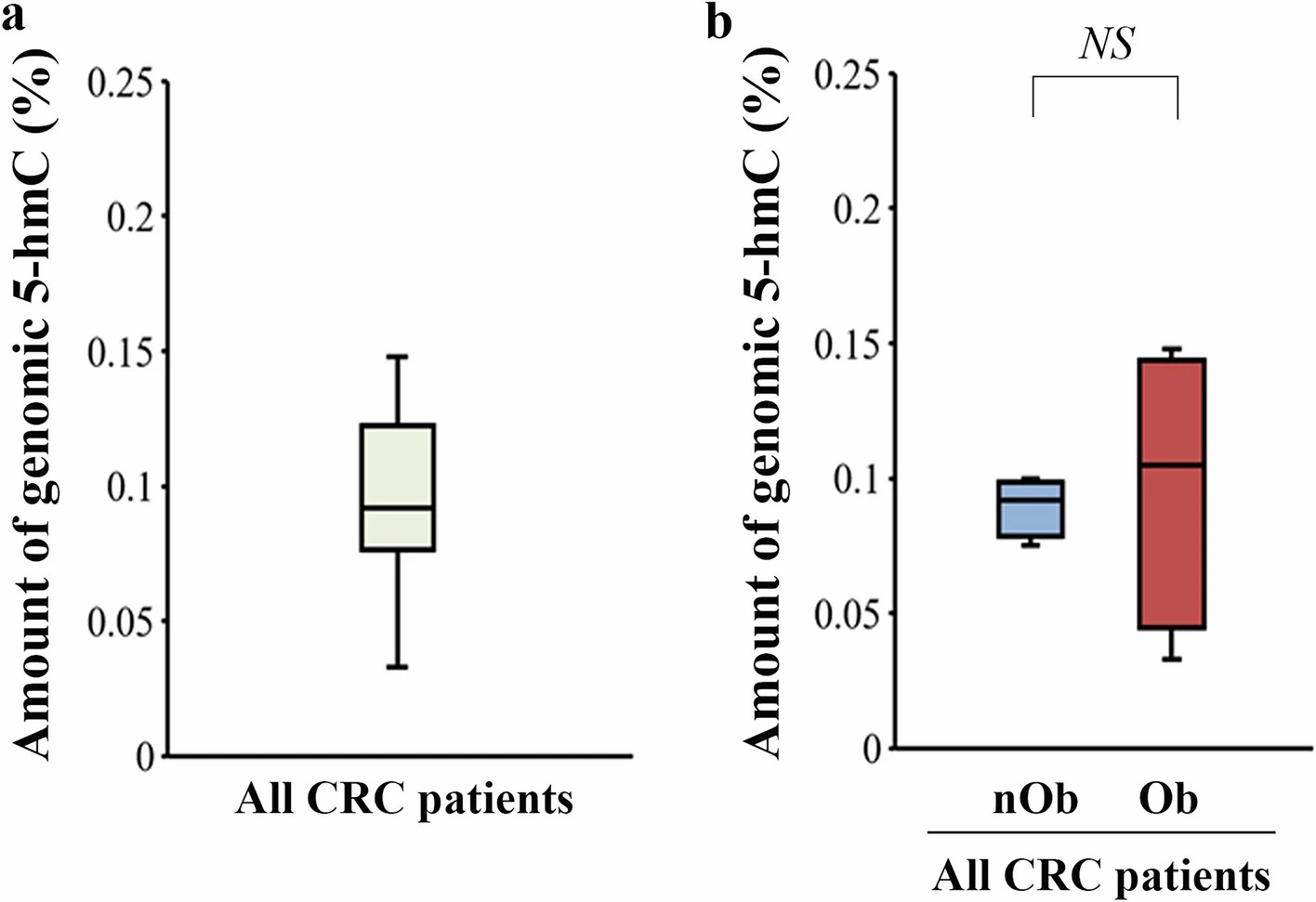
Adapted from Kon et al. (2023). Modulation of AMPK/TET2/5-hmC axis in response to metabolic alterations as a novel pathway for obesity-related colorectal cancer development. Scientific Reports 13:2858. (CC BY 4.0)
Representative Results: DNA/RNA Modification Immunoassay Data Outputs and Platform Performance
Our DNA/RNA modification immunoassay platform delivers quantitative modification data through colorimetric, chemiluminescent, fluorescent, and qPCR-based readouts, matched to the detection sensitivity and throughput requirements of each research project. The representative data below illustrates the typical output quality and platform specifications achieved across our four immunoassay formats.
Platform Performance Specifications
| Performance Parameter |
Competitive ELISA (Colorimetric) |
Dot Blot (Chemiluminescent) |
Fluorometric ELISA |
Antibody Enrichment + qPCR |
| Detection format |
96-well plate, colorimetric (450 nm) |
Nitrocellulose membrane, ECL imaging |
96-well plate, fluorescence (Ex/Em) |
Immunoprecipitation + qPCR |
| Quantification type |
Global % modification (standard curve) |
Semi-quantitative (signal/nucleic acid ratio) |
Global % modification (standard curve) |
Locus-specific enrichment (ΔΔCt) |
| Limit of detection |
0.01–0.1% modification fraction |
0.05–0.5% modification fraction |
0.003–0.03% modification fraction |
1.5–2-fold enrichment above background |
| Sample requirement |
50–200 ng DNA/RNA per well |
200 ng–1 μg per dot |
50–100 ng DNA/RNA per well |
0.5–5 μg DNA/RNA per IP |
| Throughput |
80–200 samples/week |
40–80 samples/week |
80–200 samples/week |
20–40 samples/week |
| Inter-assay precision |
<15% CV |
<20% CV |
<15% CV |
<25% CV (enrichment-dependent) |
| Sample type compatibility |
gDNA, total RNA, mRNA, cfDNA, FFPE DNA |
gDNA, total RNA, mRNA |
gDNA, total RNA, mRNA, cfDNA |
gDNA, mRNA, total RNA |
| Modification targets |
5mC, 5hmC, m⁶A, 8-oxo-dG, m⁵C, 5fC, 5caC, 6mA, N7-meG, O⁶-meG |
m⁶A, m⁵C, Ψ, m¹A, m⁷G, ac⁴C, 5mC, 5hmC |
8-oxo-dG, 8-oxo-dA, etheno-dA, low-abundance modifications |
5mC, 5hmC, m⁶A, m⁵C at defined loci |
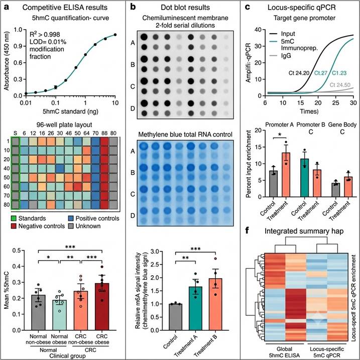
Representative data outputs from our DNA/RNA Modification Immunoassay platform. Left: Competitive ELISA standard curve and plate layout for 5hmC quantification in clinical FFPE samples. Center: Dot blot m⁶A detection with chemiluminescent imaging and densitometric quantification. Right: MeDIP-qPCR locus-specific 5mC quantification with amplification curves and percent input enrichment analysis.
Key data deliverables included in every DNA/RNA modification immunoassay project:
- ELISA quantification data package — Complete quantification results with calibration curve (four-parameter logistic regression parameters, R² value), absorbance values for all samples, standards, and controls, calculated modification percentage (%5mC, %5hmC, %m⁶A, or modification/10⁶ nucleotides for 8-oxo-dG) with intra- and inter-plate quality control metrics, and plate layout documentation
- Dot blot image and quantification package — High-resolution chemiluminescent images (TIFF format) for modification-specific antibody detection and total nucleic acid loading control (methylene blue or SYBR Gold), densitometric quantification values for all spots, normalized modification signal (modification signal intensity/loading control intensity) for each sample, and statistical comparison across experimental groups
- qPCR enrichment data package — qPCR amplification curves and Ct values for each target region across input, IP (modification antibody), and IgG control samples, percent input or enrichment fold-change calculations using the ΔΔCt method, melt curve analysis for amplicon specificity verification, and primer sequence documentation
- Quality control documentation — Antibody validation data (specificity testing, lot number, recommended dilution), standard curve performance (linear range, limit of detection, limit of quantification), positive and negative control sample results, and inter-assay reproducibility metrics across analytical batches
- Methods documentation — Complete protocols for DNA/RNA extraction, denaturation, ELISA/dot blot/qPCR-compatible detection, and data analysis, formatted for publication methods sections and regulatory reference
Related Services
Our DNA/RNA modification immunoassay platform is part of a comprehensive DNA/RNA modification analysis service portfolio spanning LC-MS quantification, modification-specific LC-MS analysis, oxidative damage assessment, adductomics, and multi-modal integration for epigenetics, epitranscriptomics, and translational research programs.
- DNA/RNA Modification LC-MS Analysis — Integrated LC-MS analysis platform covering both DNA and RNA modifications in a single analytical workflow for comprehensive nucleoside modification profiling with orthogonal validation of immunoassay findings
- RNA Modification Quantification by LC-MS/MS — Mass spectrometry-based absolute quantification of 40+ modified ribonucleosides using isotope dilution LC-MS/MS for comprehensive epitranscriptomics analysis
- m⁶A Modification LC-MS Analysis — Dedicated LC-MS analysis of N⁶-methyladenosine (m⁶A) and related adenine modifications with optimized enrichment and quantification protocols
- Oxidative DNA/RNA Damage Assay — Targeted LC-MS/MS quantification of oxidatively damaged nucleoside species in DNA and RNA for oxidative stress research and orthogonal validation of 8-oxo-dG ELISA results
- DNA/RNA Adductomics — Comprehensive LC-MS/MS analysis of DNA and RNA adducts for environmental exposure assessment, toxicology, and carcinogenesis research
- mRNA Modification LC-MS Analysis — Targeted LC-MS/MS profiling of modified ribonucleosides in purified mRNA with polyA selection and rRNA depletion workflows
FAQs
How do DNA/RNA modification immunoassays compare with LC-MS/MS quantification?
Immunoassays and LC-MS/MS are complementary approaches with distinct advantages. Immunoassays (ELISA, dot blot) offer lower cost per sample (typically $5–20/sample vs. $50–150 for LC-MS/MS), higher throughput (80–200 samples/week vs. 20–40 for LC-MS/MS), minimal sample preparation, and compatibility with standard laboratory equipment. However, immunoassays provide relative or semi-quantitative data based on antibody binding, may show cross-reactivity with structurally similar modifications, and cannot distinguish modifications at the sequence level. LC-MS/MS provides absolute molar quantification (fmol/μg RNA), unambiguous chemical identification through mass measurement and fragmentation, and simultaneous detection of multiple modifications in a single run. The choice between approaches depends on your research goals: immunoassays are ideal for high-throughput screening and large cohort studies, while LC-MS/MS is preferred for rigorous quantification, multi-modification profiling, and cross-platform validation.
What modification-specific antibodies are available for DNA/RNA modification immunoassays?
Our validated antibody panel includes monoclonal and polyclonal antibodies against 15+ DNA and RNA modifications. For DNA modifications: 5-methylcytosine (5mC, clone 33D3), 5-hydroxymethylcytosine (5hmC, clone RM236), 5-formylcytosine (5fC), 5-carboxylcytosine (5caC), N6-methyladenine (6mA), N7-methylguanine (N7-meG), O6-methylguanine (O⁶-meG), 8-oxo-7,8-dihydro-2'-deoxyguanosine (8-oxo-dG, clone 483.15), and 8-oxo-7,8-dihydro-2'-deoxyadenosine (8-oxo-dA). For RNA modifications: N6-methyladenosine (m⁶A, clones 17-3-1-1 and 202003), 5-methylcytidine (m⁵C, clone 4G3), pseudouridine (Ψ, clone 20-100-1), 1-methyladenosine (m¹A), 7-methylguanosine (m⁷G), N4-acetylcytidine (ac⁴C), inosine (I), and 8-oxo-7,8-dihydroguanosine (8-oxo-rG). Each antibody is validated for its specific immunoassay format (ELISA, dot blot, or immunoprecipitation) with documented specificity testing results.
What are the sample requirements for each immunoassay format?
Sample requirements vary by assay format and target modification. For competitive ELISA: 50–200 ng of DNA or RNA per well in duplicate, with total sample requirement of 100–400 ng per modification target. For dot blot: 200 ng–1 μg of DNA or RNA per dot, with 2–3 serial dilutions recommended for optimal quantification. For fluorometric ELISA: 50–100 ng of DNA or RNA per well, suitable for limited samples where colorimetric ELISA sensitivity is insufficient. For antibody enrichment with qPCR (MeDIP-qPCR, m⁶A-qPCR): 0.5–5 μg of fragmented DNA or RNA per immunoprecipitation reaction. For all formats, DNA/RNA should be pure (A260/A280 > 1.8 for DNA, > 2.0 for RNA), free of protein and organic solvent contamination, and quantified accurately by fluorometric method (Qubit) for precise input normalization.
Can FFPE tissue samples be used for DNA/RNA modification immunoassays?
Yes — FFPE tissue samples are compatible with all our DNA/RNA modification immunoassay platforms, with optimized DNA/RNA extraction protocols that include extended proteinase K digestion (56°C, 16–24 hours) and de-crosslinking to reverse formaldehyde-induced nucleic acid-protein crosslinks. For DNA modification analysis (5mC, 5hmC, 8-oxo-dG), FFPE-derived DNA quality is assessed by spectrophotometry and agarose gel electrophoresis before assay, with sample input adjusted for DNA fragmentation degree if necessary. For RNA modification analysis (m⁶A, m⁵C, Ψ) from FFPE tissues, RNA is typically more degraded than from fresh tissues, and we recommend using shorter amplicons (60–100 bp) for qPCR-compatible detection and increased RNA input (2–5×) for ELISA and dot blot. FFPE compatibility is a critical advantage for clinical and translational research, enabling access to the vast archives of clinically annotated tissue samples in hospital pathology departments and biobanks.
How specific are modification-specific antibodies? Can they cross-react with other modifications?
Antibody specificity is the most critical factor in immunoassay-based modification detection. Our antibodies are rigorously validated for specificity against a panel of structurally related modified and unmodified nucleosides. For example, the 5hmC antibody (clone RM236) is tested against 5mC, 5fC, 5caC, and unmodified cytosine to ensure selective 5hmC binding. However, some cross-reactivity can occur in certain conditions: for instance, some m⁶A antibodies show partial cross-reactivity with m⁶Am (2'-O-methyl-N6-methyladenosine), requiring confirmation by orthogonal methods or RNase treatments. We provide specificity characterization data for all antibodies used in our assays, recommend appropriate controls (competition controls with excess free modified nucleoside, knockout/knockdown controls where available) to validate modification specificity, and always offer optional LC-MS/MS cross-validation to confirm key findings from immunoassay-based detection.
What is the difference between global and locus-specific modification quantification?
Global modification quantification (ELISA, dot blot) measures the average modification level across all DNA or RNA in the sample, providing a single value representing the bulk modification content (e.g., %5hmC in total genomic DNA, %m⁶A in total mRNA). This is useful for assessing overall epigenetic or epitranscriptomic state, screening for modification changes across conditions, and identifying samples with extreme modification levels. Locus-specific quantification (MeDIP-qPCR, hMeDIP-qPCR, m⁶A-qPCR) measures modification enrichment at a specific genomic region or transcript, providing information about modification distribution at individual genes or regulatory elements. The choice depends on the research question: global quantification is appropriate for assessing overall epigenetic/epitranscriptomic status, while locus-specific quantification is needed for understanding modification-based regulation of specific genes or pathways. Our service can perform both global and locus-specific analysis on the same samples in an integrated workflow.
How are DNA/RNA modification immunoassay data analyzed and reported?
Data analysis and reporting depend on the immunoassay format. For ELISA: raw absorbance values are background-corrected, standard curves are fitted using four-parameter logistic regression, and sample modification percentages are interpolated from the standard curve. For dot blot: chemiluminescent images are quantified by densitometry (ImageJ or Image Lab software), modification signal is normalized to total nucleic acid loading (methylene blue or SYBR Gold staining), and normalized values are compared across groups. For qPCR-compatible detection: amplification curves are analyzed for Ct values, percent input or enrichment fold-change is calculated using the ΔΔCt method relative to input DNA/RNA or IgG control, and melt curves are inspected for amplicon specificity. Statistical analysis includes tests appropriate for the experimental design (Student's t-test, Mann-Whitney, one-way or two-way ANOVA, with appropriate post-hoc tests and multiple testing correction). Data are delivered with complete documentation including raw data files, analysis parameters, and all quality control metrics.
References
- Kon T, Umehara M, Okahashi A, et al. Modulation of AMPK/TET2/5-hmC axis in response to metabolic alterations as a novel pathway for obesity-related colorectal cancer development. Scientific Reports. 2023;13:2858.
- Ensinck I, Sideri T, Modic M, Capitanchik C, Toolan-Kerr P, van Werven FJ. m6A-ELISA, a simple method for quantifying N6-methyladenosine from mRNA populations. RNA. 2023;29(5):705–712.
- Walker NJ, Rashid M, Yu S, et al. Hydroxymethylation profile of cell-free DNA is a biomarker for early colorectal cancer. Scientific Reports. 2022;12:16566.
For research use only. Not for use in diagnostic procedures.