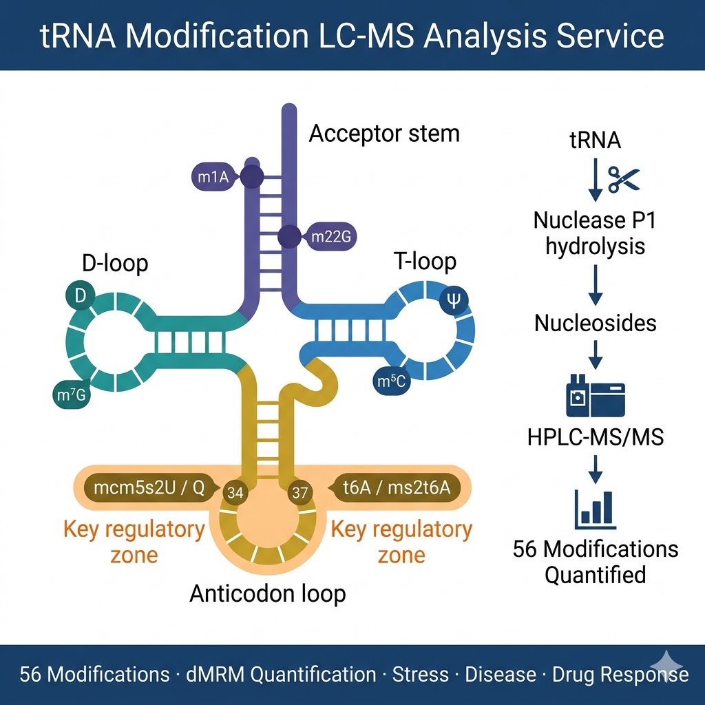
tRNA Modifications — From Structural Marks to Dynamic Regulators of Translation
Transfer RNA occupies a unique position in cell biology: it is simultaneously the most abundant RNA species in a cell (comprising ~15% of total cellular RNA), the most extensively modified (with modifications at roughly one in six nucleotide positions), and the most direct molecular link between the information content of the genome and the activity of the proteome. Every amino acid incorporation event requires a correctly modified, correctly charged tRNA — and the modifications on that tRNA directly affect how efficiently and accurately the event occurs.
Structural Modifications — Folding, Stability, and Rigidity
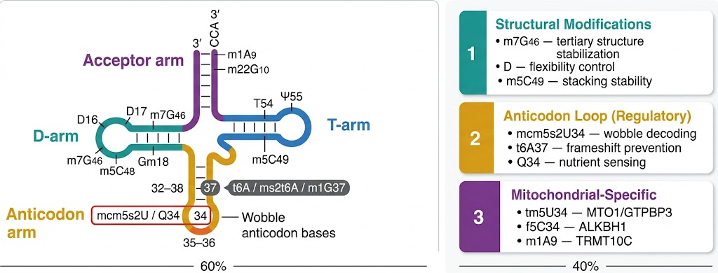
Modifications on the tRNA body (D-loop, T-loop, variable loop) primarily serve structural functions — maintaining the three-dimensional L-shaped architecture required for ribosome recognition and aminoacyl-tRNA synthetase binding. m7G46 (7-methylguanosine in the variable loop) stabilizes the tRNA tertiary structure through electrostatic interactions; m5C49 and m5C50 in the T-loop contribute to stacking stability; dihydrouridine (D) in the D-loop increases single-strand flexibility by disrupting base stacking. Loss of these modifications impairs tRNA folding and accelerates degradation via the RTD (rapid tRNA decay) pathway in eukaryotes. Mitochondrial tRNAs are particularly dependent on their modification complement for structural integrity — mutations in mitochondrial tRNA modification enzymes (TRMT2B, YRDC, GTPBP3) cause severe mitochondrial encephalomyopathies because the unmodified tRNA population is rapidly degraded.
Anticodon Loop Modifications — Decoding Fidelity and Translational Efficiency
The modifications most directly linked to translational regulation cluster in the anticodon loop, particularly at positions 34 (wobble position) and 37 (immediately 3′ of the anticodon). Position 34 modifications — including mcm5s2U34, mcm5U34, cmo5U34, ncm5U34, and queuosine (Q34) — determine the wobble-base pairing capacity of the tRNA, controlling which codons it can decode and with what efficiency. Position 37 modifications — including t6A37, ms2t6A37, i6A37, and m1G37 — reinforce the first codon-anticodon base pair and prevent translational frameshifting. The wobble modifications are installed by multi-enzyme complexes (Elongator for mcm5 side chains; methylthiotransferases for ms2; CDKAL1, KEOPS for t6A) whose defects are directly linked to diabetes (CDKAL1 mutations impair insulin codon translation), neurological disease (Elongator mutations cause familial dysautonomia), and cancer (Elongator/Elp3 expression is dysregulated in multiple cancer types).
Dynamic tRNA Modification Reprogramming — Stress Response and Disease
The discovery that tRNA modifications are dynamic — not static structural features but actively regulated in response to environmental and cellular signals — fundamentally changed our understanding of translational control. Under oxidative stress, Trm4-mediated m5C at the wobble position of tRNALeu(CAA) increases, selectively promoting translation of mRNAs enriched in TTG codons — including stress-survival proteins with codon-biased sequences. Nutrient restriction, heat shock, and genotoxic stress each produce distinct, reproducible signatures of tRNA modification change detectable by LC-MS profiling. These signatures reflect the activation of specific tRNA-modifying enzymes (or their inhibition) as part of the cellular stress response — directly coupling the epitranscriptome to translational regulation of the stress proteome. In human disease, this means that tRNA modification profiles in patient tissue or circulating cells contain diagnostic and mechanistic information about the state of cellular stress pathways, metabolic programs, and translational regulation that no other single measurement captures.
tRNA Modifications in Cancer
Multiple lines of evidence connect tRNA modification dysregulation to cancer biology. Elp3 (the catalytic subunit of the Elongator complex, responsible for mcm5U34/mcm5s2U34 modifications) is overexpressed in breast cancer and promotes metastasis through codon-biased translation of invasion-promoting proteins. NSUN2 (m5C tRNA methyltransferase) is overexpressed in multiple cancer types and protects tRNAs from angiogenin cleavage — its dysregulation alters translation efficiency and cell proliferation. ALKBH3 (demethylase for m1A and m3C) affects tRNA-mediated translation in pancreatic cancer. Queuosine (Q34) modification, which is nutrient-sensing and depends on dietary queuine availability, is reduced in cancer cells — correlating with altered decoding efficiency. LC-MS tRNA modification profiling in cancer cell models, patient-derived organoids, or tissue biopsies can identify cancer-specific modification signatures and mechanistically link tRNA modification enzyme expression changes to translational reprogramming of oncogenic codon-biased mRNAs.
Mitochondrial tRNA Modifications and Disease
Human cells contain two distinct tRNA populations: cytoplasmic (encoded by nuclear DNA) and mitochondrial (encoded by mitochondrial DNA, 22 tRNA genes). Mitochondrial tRNAs are even more dependent on post-transcriptional modifications than cytoplasmic tRNAs — they lack the extensive base-pairing and structural redundancy of cytoplasmic tRNA, making modifications essential for compensating structural limitations. The major mitochondrial tRNA modifications — τm5U34 (5-taurinomethyluridine, installed by MTO1/GTPBP3), f5C34 (5-formylcytidine, by ALKBH1), and m1A9 (by TRMT10C/SDR5C1) — are essential for mitochondrial translation. Mutations affecting these enzymes cause MELAS (mitochondrial encephalomyopathy, lactic acidosis, stroke), MERRF (myoclonic epilepsy with ragged red fibers), Leigh syndrome, and other mitochondrial disorders. LC-MS profiling of total tRNA from patient cells or tissues can distinguish cytoplasmic from mitochondrial modification defects and quantify the modification deficit at specific positions — providing mechanistic diagnostic information complementary to genetic sequencing.
Why LC-MS Is the Reference Method for tRNA Modification Analysis
Alternative approaches for tRNA modification analysis include: antibody-based detection (limited to individual modifications for which specific antibodies exist — m6A, m5C, pseudouridine; prone to cross-reactivity); sequencing-based methods (nanopore direct RNA sequencing, m5C-seq, PseU-seq — sensitive but limited to specific modification types, requiring complex bioinformatics and often providing relative rather than absolute quantification); and metabolic labeling (applicable to nascent modification dynamics but not steady-state profiling). LC-MS by nucleoside mass spectrometry is the only approach that simultaneously, quantitatively measures the full modification profile — all 56 modification types in a single analytical run — providing absolute or relative abundance data, without antibody requirements, without sequencing-level complexity, and with the analytical precision (CV typically <10% within-run, <15% between runs) required for differential analysis across biological conditions. It is the method of choice endorsed by the Suzuki, Dedon, and Helm laboratories — the field's leading methodological groups.
LC-MS tRNA Modification Analysis Platform
To detect changes in the abundance of different modifications on tRNA, Creative Proteomics offers a one-stop solution for LC-MS analysis of tRNA modification quantification. This service provides analysis of 56 types of tRNA nucleoside modifications, starting from total RNA or purified tRNA as input material.
Analytical Principle
tRNA modifications are analyzed at the nucleoside level following complete enzymatic hydrolysis of the RNA backbone. This approach provides the most comprehensive, quantitative, and reproducible measurement of tRNA modification abundance available: each modified nucleoside is detected as a discrete chromatographic peak with a characteristic parent ion → product ion transition (MRM transition) in the triple-quadrupole mass spectrometer, enabling unambiguous identification and precise quantification of each modification type simultaneously.
The nucleoside-level approach is compatible with all modification types — including labile modifications (thiolations, hypermodified bases) that are lost or altered during oligonucleotide-level MS/MS fragmentation approaches. Unlike sequencing-based methods, which require computational inference of modification identity from signal changes, nucleoside LC-MS directly measures the chemical identity and abundance of each modified nucleoside with analytical standards confirming each assignment.
Instrument Platform & Detection Method
HPLC system: Agilent 1290 Infinity II UHPLC with reversed-phase C18 column optimized for nucleoside separation. Gradient elution using aqueous ammonium acetate/methanol mobile phases resolves nucleosides by hydrophobicity and polarity — canonical nucleosides (A, G, C, U) elute first, followed by modified nucleosides in order of increasing hydrophobicity and modification mass. The optimized 30–45 min gradient resolves >56 nucleosides with baseline separation for most pairs.
Mass spectrometer: Agilent 6470 triple quadrupole (QQQ) with Agilent Jet Stream ESI source in positive ion mode. Dynamic multiple reaction monitoring (dMRM): each nucleoside is monitored by its characteristic parent ion → fragment ion (typically [M+H]+ → [nucleobase+H]+ or [ribose loss fragment]) with optimized collision energy and fragmentor voltage per analyte. Retention time windows are applied to each MRM transition to maximize dwell time and minimize cross-talk. A minimum of two MRM transitions per modification are monitored where possible to confirm identity alongside chromatographic retention time.
Quantification: peak areas are extracted for each modification using MassHunter Quantitative Analysis. Isotope-labeled internal standards (SILIS — stable isotope-labeled internal standards, biosynthetically produced or synthetic) are co-processed with samples to correct for matrix effects and extraction variability. Calibration curves are established for each modification type using authentic synthetic or biosynthetically-derived standards of confirmed purity. Results are reported as modification abundance per 106 canonical nucleosides, modification ratio to canonical A/G/C/U, or relative ratio between experimental groups.
Service Highlights
- 56 modifications in one run: the most comprehensive single-run tRNA modification panel available, covering all major anticodon loop, D-loop, T-loop, and body modifications relevant to tRNA function and disease.
- High sensitivity: attomole-level detection for high-abundance modifications (m6A, m7G, pseudouridine); low femtomole detection for rare modifications — enabling analysis from as little as 1–5 μg purified tRNA or 5–10 μg total RNA.
- Analytical precision: within-run CV <10% for high-abundance modifications; between-run CV <15%. Sufficient for differential analysis between conditions with ≥3 biological replicates per group.
- Agilent platform: optimized pre-treatment and high-quality Agilent equipment ensure comprehensive and accurate analysis of base modifications — elevating sensitivity, precision, dynamic range, and robustness to publication standard.
- Mitochondrial and cytoplasmic modification coverage: the nucleoside-level approach captures both cytoplasmic and mitochondrial tRNA modifications simultaneously from total cellular tRNA, enabling comparison of modification states across both tRNA populations in a single experiment.
- No sequencing or antibody requirements: direct chemical measurement of modification identity and abundance — unambiguous and independent of computational prediction or antibody cross-reactivity.
Types of Analysis — 56 tRNA Nucleoside Modifications Quantified
The table below lists all 56 tRNA nucleoside modifications covered by our LC-MS panel. Each modification is quantified by its characteristic parent ion → product ion MRM transition with retention time confirmation. Modifications are ordered by analysis number as in our validated method panel.
| No. |
Nucleoside |
Symbol |
No. |
Nucleoside |
Symbol |
| 1 |
3′-O-methyladenosine |
3′-OMeA |
27 |
3′-O-methyluridine |
3′-OMeU |
| 2 |
2′-O-methylcytidine |
Cm |
28 |
5-methyl-2-thiouridine |
m5s2U |
| 3 |
3-methylcytidine |
m3C |
29 |
5-methoxyuridine |
mo5U |
| 4 |
5-methylcytidine |
m5C |
30 |
pseudouridine |
Ψ |
| 5 |
N6-isopentenyladenosine |
i6A |
31 |
2′-O-methylinosine |
Im |
| 6 |
5,2′-O-dimethylcytidine |
m5Cm |
32 |
3-methyluridine |
m3U |
| 7 |
1-methyladenosine |
m1A |
33 |
1-methylpseudouridine |
m1Ψ |
| 8 |
2-thiocytidine |
s2C |
34 |
5-hydroxymethylcytidine |
hm5C |
| 9 |
N2,N2,7-trimethylguanosine |
m2,2,7G |
35 |
5,2′-O-dimethyluridine |
m5Um |
| 10 |
N4-acetyl-2′-O-methylcytidine |
ac4Cm |
36 |
N6-threonylcarbamoyladenosine |
t6A |
| 11 |
N6-methyladenosine |
m6A |
37 |
2-methylthio-N6-threonylcarbamoyladenosine |
ms2t6A |
| 12 |
3′-O-methylcytidine |
3′-OMeC |
38 |
5-carboxymethyluridine |
cm5U |
| 13 |
2′-O-methyladenosine |
Am |
39 |
5-methoxycarbonylmethyl-2-thiouridine |
mcm5s2U |
| 14 |
N2,N2-dimethylguanosine |
m22G |
40 |
5-methoxycarbonylmethyluridine |
mcm5U |
| 15 |
5′-O-methylthymidine |
5′-OMeT |
41 |
2-methylthio-N6-isopentenyladenosine |
ms2i6A |
| 16 |
2′-O-methyluridine |
Um |
42 |
Peroxywybutosine |
o2Yw |
| 17 |
inosine |
I |
43 |
5-taurinomethyl-2-thiouridine |
tm5s2U |
| 18 |
2′-O-methylguanosine |
Gm |
44 |
5-oxyacetic acid uridine |
cmo5U |
| 19 |
1-methylguanosine |
m1G |
45 |
5-carbamoylmethyluridine |
ncm5U |
| 20 |
7-methylguanosine |
m7G |
46 |
Queuosine |
Q |
| 21 |
N2-methylguanosine |
m2G |
47 |
5-taurinomethyluridine |
tm5U |
| 22 |
3′-O-methylinosine |
3′-OMeI |
48 |
5-formyl-2′-O-methylcytidine |
f5Cm |
| 23 |
2-thiouridine |
s2U |
49 |
dihydrouridine |
D |
| 24 |
4-thiouridine |
s4U |
50 |
5-formylcytidine |
f5C |
| 25 |
5-methyluridine |
m5U |
51 |
wybutosine |
yW |
| 26 |
N4-acetylcytidine |
ac4C |
52 |
5-methoxycarbonylmethyl-2′-O-methyluridine |
mcm5Um |
|
53 |
5-aminomethyl-2-thiouridine |
nm5s2U |
|
54 |
2-methylthio-N6-(cis-hydroxyisopentenyl)adenosine |
ms2io6A |
|
55 |
N6-(cis-hydroxyisopentenyl)adenosine |
io6A |
|
56 |
5-methylaminomethyl-2-selenouridine |
mnm5se2U |
All modifications are covered by validated dMRM transitions with authentic synthetic or biosynthetically-produced nucleoside standards for identity confirmation. Coverage of individual modifications depends on their abundance in the specific RNA fraction analyzed — rare modifications present at <1 per 10,000 tRNA molecules may be below detection threshold in small input samples. Contact us to confirm coverage for specific modifications of interest before project initiation.
tRNA Modification LC-MS Analysis Workflow
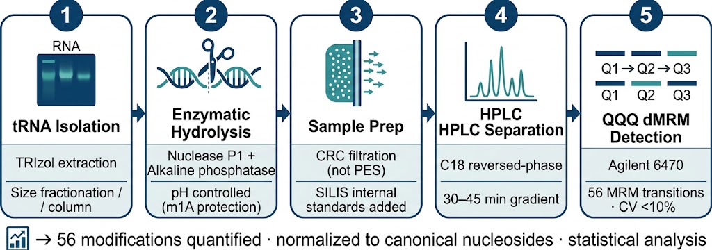
Step 1 — RNA Extraction & tRNA Isolation
Total RNA is extracted from cells or tissue using guanidinium thiocyanate-phenol-chloroform extraction (TRIzol or equivalent) with RNase-free conditions throughout. RNA integrity and concentration are assessed by Bioanalyzer or TapeStation (RIN ≥7.0 required). tRNA is isolated from total RNA by two complementary methods: (1) size fractionation by PAGE or small RNA enrichment column — isolating the small RNA fraction (<200 nt) containing tRNA, 5S rRNA, and small ncRNAs; and (2) tRNA-specific affinity chromatography for projects requiring highly purified tRNA free from other small RNA species. The tRNA fraction is quantified by Qubit fluorometry and an aliquot retained for quality assessment. All RNA handling is performed in RNase-free conditions with DEPC-treated water and RNase inhibitors.
Step 2 — Enzymatic Hydrolysis to Nucleosides
Purified tRNA is completely digested to individual nucleosides using a two-enzyme system: Nuclease P1 (from Penicillium citrinum; 40 mU per 10 μg tRNA) in sodium acetate buffer pH 5.3 with ZnCl2, incubated at 37 °C for 1.5–2 h, followed by alkaline phosphatase (shrimp alkaline phosphatase; 0.1 U) after pH adjustment to remove 5′-phosphate groups. The complete reaction generates individual nucleosides with free 3′-OH and 5′-OH groups, ready for HPLC injection. Hydrolysis completeness is verified by the absence of remaining RNA by UV absorbance (A260) after precipitation. Important: 1-methyladenosine (m1A) undergoes Dimroth rearrangement to N6-methyladenosine (m6A) at alkaline pH — we maintain reaction conditions at near-neutral pH and minimize incubation time at pH >7 to prevent this artifact, which would otherwise cause m1A underquantification and m6A false-positive signal.
Step 3 — Sample Cleanup & Internal Standard Addition
Hydrolyzed nucleoside mixtures are filtered to remove enzyme proteins and RNA fragments — using validated composite regenerated cellulose (CRC) filtration membranes, not polyethersulfone (PES) filters, which have been documented to cause loss of hydrophobic modified nucleosides including i6A, ms2i6A, and o2Yw. Isotope-labeled internal standards (biosynthetically-produced SILIS or synthetic isotope-labeled nucleosides where available) are added at defined concentrations before HPLC injection to account for matrix effects and injection variability. Samples are stored at −80 °C until LC-MS analysis; thiolated modifications (s4U, s2U, mcm5s2U, tm5s2U) are particularly susceptible to oxidation and are analyzed within 24 h of hydrolysis.
Step 4 — Reversed-Phase HPLC Separation
Nucleosides are separated by reversed-phase HPLC on an Agilent Poroshell 120 EC-C18 or equivalent column (2.1 mm × 50 mm, 1.9 μm, 40 °C column temperature) using a binary gradient of aqueous mobile phase A (5 mM ammonium acetate pH 5.3, 0.1% formic acid) and organic mobile phase B (methanol or acetonitrile). The gradient begins at 0% B, gradually increases to 40–60% B over 30–45 min, then returns to initial conditions. This gradient resolves canonical nucleosides from each other and from all 56 modified nucleosides by polarity, enabling retention time-based identity confirmation that is independent of — and orthogonal to — the MRM mass-based identification. UV detection at 254 nm provides an additional orthogonal confirmation layer for abundant modifications.
Step 5 — Triple Quadrupole MS/MS Detection (dMRM)
Separated nucleosides are detected by positive ion electrospray ionization (ESI) triple quadrupole mass spectrometry in dMRM mode. Each modification is monitored by a pre-validated parent ion → product ion transition: the typical fragmentation pathway is [M+H]+ (protonated molecular ion) → [nucleobase + H]+ (nucleobase loss from the ribose via glycosidic bond cleavage) or [ribose-loss fragment]. Collision energy and fragmentor voltage are individually optimized per modification using the Agilent MassHunter Optimizer. Time-scheduled MRM windows restrict monitoring of each transition to the expected elution window, maximizing dwell time per analyte and minimizing cross-talk between co-eluting species. At minimum two MRM transitions are acquired per modification where structurally possible — a primary quantifier and a secondary qualifier — to confirm analyte identity.
Step 6 — Data Analysis & Reporting
Peak areas are extracted from dMRM chromatograms using Agilent MassHunter Quantitative Analysis. Modification abundance is normalized to the sum of canonical nucleoside (A + G + C + U) peak areas — expressing each modification as modifications per 106 canonical nucleosides (or per 1,000 tRNA molecules, where tRNA molecular weight is known). Between-condition fold-changes are calculated and tested by t-test with Benjamini-Hochberg FDR correction across all 56 modifications simultaneously. Hierarchical clustering and principal component analysis of the full modification profile across samples visualizes group separation and modification co-regulation patterns. Deliverables: raw LC-MS data files, modification quantification table (all 56 modifications × all samples), normalized abundance matrix, differential analysis results (fold-change, p-value, adjusted p-value), heatmap, PCA plot, and comprehensive project report with publication-ready methods text.
Sample Requirements
| Starting Material |
Minimum Input |
Key Requirements |
| Total RNA |
≥5 μg (standard); ≥2 μg (low-input) |
RIN ≥7.0; prepared with RNase-free reagents; store at −80 °C; ship on dry ice with RNase inhibitors. Provide A260/A280 ≥1.9 and A260/A230 ≥2.0 confirmation |
| Purified tRNA fraction |
≥1 μg (standard); ≥500 ng (low-input) |
Isolated by PAGE or column chromatography; free of rRNA contamination; A260/A280 ≥1.9; store at −80 °C; ship on dry ice in RNase-free water or TE buffer |
| Cultured cells (for RNA extraction in-house) |
≥2 × 10⁶ cells per sample |
Harvest cells rapidly — do not allow RNA degradation; wash 2× with cold PBS; snap-freeze pellet in liquid nitrogen immediately; store at −80 °C; ship on dry ice |
| Tissue |
≥20 mg fresh/frozen tissue |
Snap-freeze in liquid nitrogen within 30 s of collection; store at −80 °C; ship on dry ice. FFPE tissue is not recommended — formalin crosslinking degrades RNA and introduces modification artifacts |
| Biological replicates |
≥3 per condition (minimum) |
Required for statistical differential analysis. For discovery projects profiling modification landscape without comparison: ≥2 replicates recommended for reproducibility confirmation |
Demo Results — Representative tRNA Modification Profiling Data
Below are representative data outputs from a typical tRNA modification LC-MS project. Each figure type addresses a distinct analytical question and is delivered in publication-ready format.
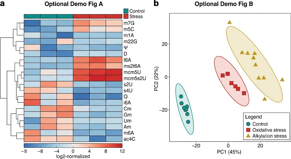
Modification Heatmap
Hierarchical clustering of 56 tRNA modification abundances across experimental conditions. Rows represent individual modification types; columns represent biological replicates. Color scale from blue (low abundance) to orange (high abundance). Key modifications: wobble modifications (mcm5U, mcm5s2U, t6A) in anticodon cluster, structural modifications (m7G, m5C, D) in body cluster.
Differential Abundance
Volcano plot comparing modification abundance between stress and control conditions. Log2 fold change on x-axis, −log10 p-value on y-axis. Significantly altered modifications highlighted: upregulated in red (mcm5U, cmo5U, t6A), downregulated in blue (m1A, m3C, ac4C). Significance cutoff: padj < 0.05, |FC| > 1.3.
PCA & Multivariate Analysis
Principal component analysis scores plot of the full 56-modification profile. Control and stress condition groups separate along PC1 (>40% variance), demonstrating condition-specific modification reprogramming. 95% confidence ellipses per group. Key loading vectors identify modifications driving group separation.
Case Study — Quantitative LC-MS of tRNA Modifications Reveals Stress-Specific, Dose-Dependent Reprogramming of the tRNA Modification Landscape
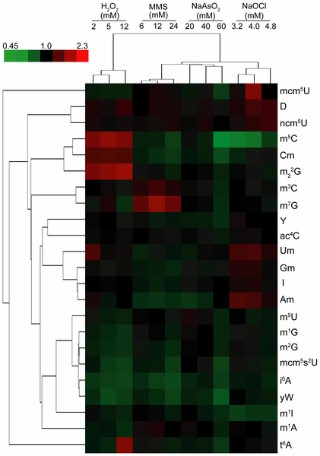
Reference: Chan CTY, Dyavaiah M, DeMott MS, Taghizadeh K, Dedon PC, Begley TJ. A quantitative systems approach reveals dynamic control of tRNA modifications during cellular stress. PLoS Genet. 2010;6(12):e1001247. DOI: 10.1371/journal.pgen.1001247 (CC BY 4.0, PMC3002981)
Background & Scientific Question
It had long been assumed that tRNA modifications were static, constitutively-installed structural features — set at the time of tRNA biogenesis and unchanged throughout the tRNA lifetime. The Begley and Dedon laboratories at MIT hypothesized that this assumption was incorrect: that tRNA modifications were in fact dynamic, condition-responsive, and that changes in their abundance in response to cellular stress could provide a systems-level regulatory signal connecting the epitranscriptome to selective translational control of stress-response proteins. To test this, they needed a method capable of quantifying the full complement of tRNA modifications in an organism — not just one or two modifications, but the complete modification profile — under multiple different stress conditions and doses. This led to the development of the first comprehensive LC-MS/MS quantitative tRNA modification profiling platform, which is the direct methodological ancestor of the service we provide today.
Methods
Cytoplasmic tRNA was isolated from Saccharomyces cerevisiae exposed to four mechanistically distinct toxicants — hydrogen peroxide (oxidative stress), arsenite (oxidative/thiol stress), methylmethanesulfonate (MMS, SN2 alkylating agent), and hypochlorite (electrophilic/chlorinating stress) — each at multiple doses. Purified tRNA was enzymatically hydrolyzed with Nuclease P1 and alkaline phosphatase to yield individual nucleosides. Modified nucleosides were separated by reversed-phase HPLC and quantified by LC-MS/MS using dynamic multiple reaction monitoring (dMRM) on a triple-quadrupole mass spectrometer. 23 of the ~25 known yeast tRNA modifications were simultaneously quantified. Multivariate statistical analysis (hierarchical clustering, principal component analysis, discriminant analysis) of the modification profiles revealed toxicant- and dose-specific patterns. Yeast mutants lacking individual tRNA modification enzymes were also profiled to identify biosynthetic dependencies and compensatory regulation between modification pathways.
Key Results
tRNA modifications are dynamic: all four stressors produced significant, reproducible changes in the abundance of multiple tRNA modifications — definitively establishing that tRNA modification profiles are not static but actively regulated in response to environmental stress.
Stress signatures are agent-specific and dose-dependent: hierarchical clustering of modification profiles correctly segregated samples by toxicant agent (>80% predictive accuracy) and by dose — demonstrating that the tRNA modification landscape encodes information about the nature and severity of cellular stress that is distinguishable by LC-MS profiling.
Modifications in the anticodon loop respond most dynamically: wobble modifications (mcm5U, mcm5s2U, cmo5U) and adjacent position 37 modifications (t6A, ms2t6A) showed the largest fold-changes under stress — consistent with their role in codon-biased translational control of stress-response mRNAs.
Cross-modification regulatory networks: profiling yeast mutants lacking individual modification enzymes revealed biosynthetic dependencies — where loss of one modification enzyme altered the abundance of modifications installed by other enzymes, revealing cross-pathway regulatory interactions not previously known.
Relevance to Our Service
This landmark study established the conceptual and technical framework for quantitative LC-MS tRNA modification profiling — demonstrating that a global, simultaneous measurement of the tRNA modification landscape across 20+ modification types reveals biologically interpretable, condition-specific regulatory signals that cannot be captured by single-modification analysis approaches. The five-stage analytical pipeline described in this paper — tRNA purification, enzymatic hydrolysis, HPLC separation, LC-MS/MS dMRM quantification, and multivariate statistical analysis — is directly implemented in our tRNA Modification LC-MS Analysis Service, extended to cover 56 modifications in the human system.
The key biological insight the study enabled — that tRNA modifications are reprogrammed as part of the stress response to facilitate selective translation of codon-biased mRNAs — is now recognized as a fundamental mechanism of translational regulation relevant to cancer, neurodegeneration, and metabolic disease. Our service provides the same quantitative modification profiling capability for your biological system of interest, whether that is a human cancer cell line, patient tissue, a model organism, or a stress treatment experiment designed to map the dynamic tRNA modification response.
References
- Chan CTY, Dyavaiah M, DeMott MS, Taghizadeh K, Dedon PC, Begley TJ. A quantitative systems approach reveals dynamic control of tRNA modifications during cellular stress. PLoS Genet. 2010;6(12):e1001247. doi.org/10.1371/journal.pgen.1001247
- Suzuki T. The expanding world of tRNA modifications and their disease relevance. Nat Rev Mol Cell Biol. 2021;22(6):375-392. doi.org/10.1038/s41580-021-00342-0
- Kellner S, Helm M, Höbartner C. Pitfalls in RNA modification quantification using nucleoside mass spectrometry. Acc Chem Res. 2023;56(23):3455-3465. doi.org/10.1021/acs.accounts.3c00402
- Nedialkova DD, Leidel SA. Optimization of codon translation rates via tRNA modifications maintains proteome integrity. Cell. 2015;161(7):1606-1618. doi.org/10.1016/j.cell.2015.05.022
FAQs — tRNA Modification LC-MS Analysis
What is the difference between tRNA modification LC-MS analysis and m6A sequencing or bisulfite-seq for m5C?
These approaches address completely different questions. m6A sequencing (m6A-seq, MeRIP-seq, m6A-CLIP) and bisulfite sequencing (BS-seq) map the position of a single modification type (m6A or m5C respectively) within individual RNA molecules at specific sequence positions — providing transcriptome-wide site-specific information about one modification. Our tRNA Modification LC-MS Analysis measures the global abundance of up to 56 different modification types as nucleosides from the bulk tRNA population — providing modification abundance (how much of each modification type is present in total) across the full chemical diversity of tRNA modifications, but without position-specific or transcript-specific resolution. These are complementary, not competing, approaches. LC-MS nucleoside profiling answers "which modification types change in abundance, and by how much, under this condition?" — which is the right first question when characterizing tRNA modification dynamics in stress, disease, or drug treatment studies. Position-specific sequencing methods then confirm which tRNA species and positions are affected. We recommend starting with LC-MS profiling for global modification landscape analysis; position-specific methods follow once priority modification types are identified.
Can you detect both cytoplasmic and mitochondrial tRNA modifications from the same sample?
Yes. When total cellular tRNA is isolated without subcellular fractionation, both cytoplasmic and mitochondrial tRNA are present in the preparation — typically in a ratio reflecting their relative abundance (~95% cytoplasmic, ~5% mitochondrial in actively proliferating cells). Our LC-MS analysis measures the total pool of each modified nucleoside, so the signal reflects the combined cytoplasmic and mitochondrial tRNA modification content. For modifications that occur predominantly in mitochondrial tRNAs (e.g., τm5U34 / tm5U — installed by MTO1/GTPBP3 in mitochondria, absent from cytoplasmic tRNAs; f5C34 — installed by ALKBH1 in mitochondria), changes in LC-MS signal are dominated by the mitochondrial tRNA component even though it constitutes only a small fraction of total tRNA by mass — because these modifications are mitochondria-specific markers. For modifications occurring in both pools (m1A, pseudouridine, m7G), the LC-MS signal represents the sum of both pools. If specific attribution to cytoplasmic vs. mitochondrial tRNA is required, subcellular fractionation followed by separate tRNA isolation from cytoplasmic and mitochondrial fractions before LC-MS analysis is available as an add-on service.
Are there any modifications in the panel that are prone to artifacts during sample preparation?
Yes — several modifications require specific precautions. 1-methyladenosine (m1A): undergoes Dimroth rearrangement to N6-methyladenosine (m6A) at mildly alkaline pH or elevated temperature — we control reaction pH to prevent this, but tRNA samples should not be stored at pH >7 or heated before hydrolysis. Thiolated modifications (s4U, s2U, mcm5s2U, tm5s2U, m5s2U): susceptible to oxidation of the thiol group — samples containing these modifications should be processed rapidly after hydrolysis and stored at −80 °C under argon where possible; we analyze thiolated modification samples within 24 h of hydrolysis. i6A and ms2i6A: can be lost by adsorption to polyethersulfone (PES) filtration membranes — we use composite regenerated cellulose (CRC) filtration exclusively. 4-thiouridine (s4U): dimerizes under UV exposure — RNA samples should be handled in subdued light. 3-methylcytidine (m3C): can deaminate to m3U under prolonged alkaline hydrolysis conditions — we limit alkaline phosphatase incubation time and pH exposure. We document all precautions applied for each project and include artifact-prone modifications with flagged QC notes in the final report.
How many biological replicates are needed, and what statistical analysis is performed?
A minimum of 3 biological replicates per condition is required for any differential analysis between conditions. With n=3, t-test or Wilcoxon signed-rank test can detect modifications with consistent fold-changes ≥1.5 and low within-group variance (CV <20%). For studies aiming to detect smaller fold-changes (<1.3-fold) or with higher biological variability, n=4–5 per condition substantially increases statistical power. Because 56 modifications are tested simultaneously, we apply Benjamini-Hochberg FDR correction to all p-values, and report both raw p-values and adjusted p-values (padj). We consider padj ≤0.05 and |FC| ≥1.3 (or user-specified thresholds) as the default significance criteria for differential modification analysis. Beyond per-modification statistical testing, we perform multivariate analysis — hierarchical clustering (Ward's method, Euclidean distance on log-transformed abundances) and PCA of the full 56-modification profile — to assess overall group separation and identify modification types that co-vary across samples. This multivariate approach is often more sensitive than per-modification t-tests for detecting condition-dependent modification reprogramming patterns, particularly when multiple modifications change in the same direction under a given condition.
Can this service be combined with mRNA sequencing, proteomics, or other omic analyses to study translational regulation?
Yes — and this type of multi-omic integration is increasingly the standard for mechanistic studies of tRNA modification-dependent translational regulation. Our tRNA Modification LC-MS Analysis is designed to be integrated with complementary analyses from the same biological samples or experiment. Most commonly this includes: (1) total RNA-seq or ribosome profiling (Ribo-seq) to measure codon-specific translation efficiency — enabling direct testing of whether changes in wobble modification abundance (e.g., mcm5s2U34 levels measured by LC-MS) correlate with changes in decoding efficiency of the corresponding codons (e.g., UUG codons in the case of tRNALeu(CAA)); (2) total proteomics or TMT proteomics to measure protein-level translational output — identifying whether the proteins whose translation changes under stress conditions are enriched for codons decoded by the modifications that change (codon-biased translation testing); (3) mRNA modification profiling by our mRNA Modification LC-MS Analysis Service or m6A-seq — to distinguish tRNA from mRNA modification contributions to translational regulation; and (4) DNA/RNA modification immunoassay via our DNA/RNA Modification Immunoassay Services for rapid, high-throughput screening of modification levels across large sample sets before LC-MS quantification. Contact us to discuss multi-omic project design and integration analysis.