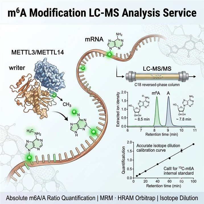
Precise and Quantitative m6A Analysis by LC-MS/MS
N6-methyladenosine (m6A) represents the most abundant internal RNA modification in eukaryotic mRNA, with approximately 25–50% of mammalian mRNAs containing at least one m6A site — typically enriched near stop codons, in 3′ UTRs, and within long internal exons. The m6A mark is installed co-transcriptionally by the METTL3/METTL14 methyltransferase complex (writers), removed by the demethylases FTO and ALKBH5 (erasers), and recognized by YTH domain-containing proteins and IGF2BP family members (readers) that mediate downstream effects on RNA fate. This dynamic regulation enables m6A to function as a versatile epitranscriptomic signal that modulates RNA splicing, nuclear export, translation initiation and efficiency, and RNA stability in response to cellular signaling, environmental stimuli, and developmental cues.
Why LC-MS/MS is the Gold Standard for m6A Quantification
While antibody-based methods (MeRIP-seq/m6A-seq) and sequencing-based approaches provide transcriptome-wide m6A distribution maps, they are subject to antibody cross-reactivity, amplification bias, and variable enrichment efficiency that can lead to false-positive assignments and imprecise quantification. Liquid chromatography-tandem mass spectrometry (LC-MS/MS) overcomes these limitations by providing direct, absolute quantification of m6A at the nucleoside level, independent of antibody quality or sequencing artifacts. Our LC-MS platform delivers accurate m6A/A ratios through isotope dilution with stable isotope-labeled internal standards (13C-m6A, 15N-A), giving researchers the quantitative precision needed to detect subtle but biologically significant changes in m6A abundance across experimental conditions. For researchers combining m6A quantification with broader RNA modification analysis, our RNA Modification LC-MS/MS service provides comprehensive epitranscriptome profiling across 50+ modified ribonucleosides. For DNA methylation analysis, our DNA Base Modification Quantification LC-MS service offers parallel quantification of 5mC, 5hmC, and related epigenetic DNA modifications.
Our m6A LC-MS Analysis Service Portfolio
We offer a structured portfolio of m6A analysis services designed to address specific research objectives — from global m6A/A ratio quantification in purified RNA fractions to multi-modification co-profiling and integration with sequencing-based m6A mapping. The table below maps common research goals to our recommended service modules.
| Research Objective |
Recommended Service |
Key Technology |
| Global m6A/A ratio quantification in mRNA |
mRNA m6A Absolute Quantification |
Poly(A) selection, enzymatic hydrolysis, LC-MS/MS with isotope dilution (13C-m6A), MRM detection |
| m6A profiling in total RNA (multi-modality) |
Total RNA m6A Quantification |
Total RNA extraction, nuclease P1/alkaline phosphatase digestion, HRAM full-scan + MRM quantification |
| m6A/m6Am/m1A co-detection and comparison |
Multi-Modification m6A Panel |
Scheduled MRM acquisition, 3-modification multiplex panel, isotopic internal standards for each analyte |
| m6A quantification in small RNA / tRNA |
Small RNA/tRNA m6A Analysis |
Size-exclusion or acid-phenol tRNA isolation, RNase T1/A digestion, LC-MS/MS with modified nucleoside library |
| Parallel LC-MS and sequencing m6A analysis |
Integrated m6A Validation Package |
LC-MS absolute quantification + MeRIP-seq/Nanopore dRNA-seq for site mapping, cross-platform correlation analysis |
| High-throughput m6A screening (clinical cohorts) |
High-Throughput m6A Quantification |
Automated sample preparation, 96-well plate format, fast LC gradient (6 min/sample), scheduled MRM acquisition |
m6A Detection Parameters and Performance Characteristics
The following table summarizes key detection parameters for m6A and related adenosine modifications quantified by our LC-MS platform. Detection limits are validated for each sample matrix and reported with batch-specific QC metrics.
| Modification |
Precursor Ion (m/z) |
Product Ion (m/z) |
Typical RT (min) |
Detection Mode |
Representative LOD |
| N6-Methyladenosine (m6A) |
282.1 [M+H]+ |
150.1 (Base) |
8.5 |
MRM (QQQ) / HRAM (Orbitrap) |
0.01% of A (5 fmol on-column) |
| Adenosine (A) |
268.1 [M+H]+ |
136.1 (Base) |
7.8 |
MRM (QQQ) / HRAM |
— (reference nucleoside) |
| N6,2′-O-Dimethyladenosine (m6Am) |
296.1 [M+H]+ |
150.1 (Base-CH3) |
9.2 |
MRM (QQQ) / HRAM |
0.01% of A |
| N1-Methyladenosine (m1A) |
282.1 [M+H]+ |
150.1 (Base) |
6.1 |
MRM (QQQ) / HRAM |
0.005% of A |
| 2′-O-Methyladenosine (Am) |
282.1 [M+H]+ |
136.1 (Base) |
9.5 |
HRAM (Orbitrap) / MRM |
0.05% of A |
| N6-Methyladenosine-13C (IS) |
288.1 [M+H]+ |
156.1 (Base) |
8.5 |
MRM (QQQ) |
— (internal standard) |
For studies requiring broader epitranscriptomic coverage beyond adenosine modifications, our mRNA and tRNA modification LC-MS services provide comprehensive profiling across 50+ modified ribonucleosides from purified RNA fractions.
Integrated LC-MS/MS Platform for m6A Quantification
Reliable m6A quantification demands careful optimization across every stage of the analytical pipeline — from RNA extraction and enzymatic digestion through high-resolution LC-MS acquisition to data processing and stoichiometry calculation. Our platform integrates established best practices and rigorous quality control measures to ensure accurate, reproducible m6A measurement across diverse sample types and experimental conditions.
RNA Extraction and Preparation
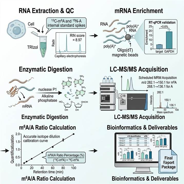
Total RNA is extracted using TRIzol-based or column-based methods optimized for RNA integrity and modification preservation. For mRNA-specific m6A quantification, poly(A)-selected mRNA is purified using oligo(dT) magnetic beads, with ribosomal RNA depletion as an alternative for lncRNA and total RNA analysis. RNA quantity and purity are assessed by UV spectrophotometry (A260/A280 ≥ 2.0), and RNA integrity is verified by capillary electrophoresis (RIN ≥ 7 for mRNA analysis). For tRNA-specific analysis, tRNA is isolated using size-exclusion chromatography or acid-phenol extraction methods. To control for recovery and matrix effects throughout the workflow, 13C- or 15N-labeled internal standards are spiked into each sample at the earliest possible step.
Enzymatic Hydrolysis and LC-MS/MS Acquisition
Purified RNA is digested to individual nucleosides using a sequential enzymatic hydrolysis protocol: nuclease P1 (37°C, 16 h) followed by alkaline phosphatase and nucleoside phosphorylase in ammonium bicarbonate buffer (37°C, 2 h). Digestion efficiency is monitored by UV absorbance and confirmed by the absence of oligonucleotide peaks in LC-MS chromatograms. Hydrolyzed nucleosides are separated by reversed-phase liquid chromatography (C18 column, 2.1 × 100 mm, 1.7 µm) using a gradient of water and acetonitrile with 0.1% formic acid. Detection is performed on triple quadrupole (QQQ) systems operating in positive ion MRM mode using mass transitions specific to m6A, A, m6Am, and m1A, or on Orbitrap HRAM systems for full-scan discovery with accurate mass confirmation.
Quantification and Normalization
m6A levels are expressed as the m6A/A ratio (%) — calculated as the peak area of m6A divided by the peak area of adenosine, normalized to internal standard recovery and calibrated against authentic m6A and A standards. Absolute quantification is performed using external calibration curves prepared in matrix-matched solutions across 4 orders of dynamic range. Method validation includes assessment of linearity (R² ≥ 0.99), accuracy (spike recovery 85–115%), precision (intra- and inter-batch CV ≤ 15%), limit of detection (S/N ≥ 3), and limit of quantification (S/N ≥ 10).
Quality Control Framework
Each analytical batch includes blank injections to monitor carryover, system suitability standards to verify instrument performance, calibration verification standards at low, medium, and high m6A/A levels, independent quality control samples from pooled RNA references, and replicate analysis of a subset of samples for inter-batch reproducibility assessment. For integrated studies combining LC-MS with sequencing-based m6A mapping, we provide cross-platform correlation analysis and orthogonal validation reports.
m6A LC-MS Analysis Workflow: From Sample to Publication-Ready Data
Step 1: RNA Extraction and Quality Control
Total RNA or mRNA is extracted using modification-preserving protocols optimized for each sample type (cells, tissues, biofluids, FFPE). RNA quantity and quality are assessed by UV spectrophotometry, fluorometric quantitation, and capillary electrophoresis (RIN score). 13C- or 15N-labeled internal standards are spiked at lysis to control for recovery.
Step 2: mRNA Enrichment (if required)
For mRNA-specific m6A analysis, poly(A)+ RNA is purified using oligo(dT) magnetic bead-based enrichment. rRNA-depleted total RNA is prepared as an alternative for transcriptome-wide analysis. Purification efficiency is verified by RT-qPCR (GAPDH enrichment ≥ 100-fold, rRNA depletion ≥ 95%).
Step 3: Enzymatic Digestion to Nucleosides
Purified RNA is sequentially digested with nuclease P1 (single-strand-specific endonuclease) and alkaline phosphatase to release individual ribonucleosides. Digestion is performed under controlled pH and temperature to prevent modification degradation. Completion is verified by UV profile.
Step 4: LC-MS/MS Acquisition
Nucleosides are separated by C18 reversed-phase LC with optimized gradient elution. Detection uses QQQ MRM (targeted quantification) or Orbitrap HRAM (full-scan profiling). Scheduled MRM with 3–5 transitions per analyte ensures specificity. 13C/15N isotope dilution corrects for matrix effects.
Step 5: m6A/A Ratio Calculation and Data Validation
m6A/A ratio (%) is calculated from isotope dilution-normalized peak areas against authentic calibration curves. Results are validated against QC acceptance criteria (linearity, accuracy, precision, LOD/LOQ). Cross-platform correlation is provided for integrated sequencing studies.
Step 6: Bioinformatics and Deliverables
Complete dataset including m6A/A ratio table with individual sample values, group statistics, and inter-batch reproducibility metrics; modification co-detection summary (when multiplexed); cross-platform correlation plots (when integrated with sequencing); and a scientist consultation session for biological interpretation.
m6A LC-MS Analysis in Biomedical Research
Quantitative m6A analysis by LC-MS supports a broad spectrum of research applications, reflecting the fundamental regulatory roles of m6A in gene expression, development, and disease. Our platform is configured to deliver the quantitative precision required for each application domain, from global m6A/A ratio measurements in model systems to clinical cohort screening and integrated multi-omics studies.
Cancer Epitranscriptomics
Dysregulation of the m6A epitranscriptome is a hallmark of multiple cancer types, with altered m6A levels driven by METTL3/METTL14 overexpression or FTO/ALKBH5 downregulation in acute myeloid leukemia, breast cancer, glioblastoma, hepatocellular carcinoma, and lung cancer. Our LC-MS platform provides the absolute m6A/A ratio quantification needed to characterize global epitranscriptomic changes in tumor versus normal tissues, monitor response to targeted therapies, and validate m6A-related biomarker candidates from sequencing-based discovery studies.
Stem Cell and Developmental Biology
m6A modification plays essential roles in embryonic stem cell self-renewal, differentiation, and lineage specification through regulation of pluripotency factor expression and developmental gene activation. Quantitative m6A profiling by LC-MS enables precise measurement of epitranscriptomic changes during stem cell differentiation, organoid development, and in vivo developmental time courses, providing the stoichiometric data needed to distinguish regulatory m6A changes from transcript abundance-driven effects.
Viral Infection and Host-Pathogen Interactions
The role of m6A in viral RNA biology — including replication, translation, and immune evasion — represents a rapidly evolving research area. Our LC-MS platform provides the orthogonal quantitative validation essential for distinguishing genuine viral RNA m6A signals from antibody-dependent artifacts, as demonstrated by recent studies using LC-MS to resolve controversies about m6A prevalence in viral genomes. The ability to quantify absolute m6A levels in purified viral RNA fractions enables rigorous testing of m6A functional hypotheses in virology research. For comprehensive nucleic acid damage and modification assessment in infection and inflammatory models, our DNA/RNA Adductomics and Damage Analysis service provides parallel modification profiling from the same sample set.
Neurological and Metabolic Disease Research
m6A modification regulates key genes in neurodevelopment, synaptic plasticity, and metabolic signaling, with m6A dysregulation linked to autism spectrum disorders, depression, obesity, and type 2 diabetes. LC-MS-based m6A quantification from brain tissue, neuronal cultures, or metabolic tissue biopsies provides the analytical sensitivity needed to detect modification changes in disease models and therapeutic intervention studies, complementing transcriptomic and proteomic datasets for multi-omics integration. For studies investigating oxidative stress contributions to disease pathology, our Oxidative DNA/RNA Damage Assay service provides parallel quantification of oxidative nucleic acid damage markers from matched samples.
Case Study: LC-MS/MS Reveals That m6A Modification Is Not a General Trait of Viral RNA Genomes
A 2024 study by Baquero-Pérez et al. published in Nature Communications applied LC-MS/MS quantification alongside orthogonal antibody-independent sequencing methods to resolve a critical controversy about whether N6-methyladenosine (m6A) is a general feature of cytoplasmic RNA virus genomes. This work highlights the essential role of LC-MS-based absolute quantification in validating RNA modification claims and establishing rigorous standards for epitranscriptomic research.
Background: Previous studies using m6A-seq (antibody-dependent immunoprecipitation) had reported extensive m6A modification across multiple cytoplasmic RNA virus genomes, including chikungunya virus (CHIKV) and dengue virus (DENV), suggesting that m6A is a general and functionally important feature of viral RNA biology. However, antibody-dependent methods are known to produce false-positive signals through non-specific binding and cross-reactivity, raising questions about whether these reported m6A sites were genuine.
Approach: The team applied a multi-method orthogonal strategy combining LC-MS/MS absolute quantification with antibody-independent techniques — including SELECT (single-base resolution) and nanopore direct RNA sequencing — to systematically evaluate m6A modification in CHIKV and DENV RNA. LC-MS/MS quantification was performed on poly(A)-selected RNA from mock-infected and virus-infected HEK293T and Huh7 cells, with m6A/A ratios determined using isotope dilution with synthetic m6A and A standards. Parallel m6A-seq experiments were conducted to directly compare antibody-dependent and antibody-independent results.
Key Findings:
- LC-MS/MS quantification showed that m6A/A ratios in poly(A)+ RNA from CHIKV- and DENV-infected cells were indistinguishable from mock-infected controls, demonstrating no measurable m6A enrichment on viral RNA
- SELECT and nanopore direct RNA sequencing independently confirmed the absence of m6A modification on CHIKV and DENV genomic RNA, consistent with LC-MS findings
- m6A-seq (antibody-dependent) produced false-positive signals in viral RNA that were not reproducible by any antibody-independent method, demonstrating that prior reports of m6A in CHIKV/DENV were artifacts of antibody cross-reactivity
- Depletion of host m6A writer (METTL3) or eraser (FTO, ALKBH5) components did not affect CHIKV or DENV viral replication
- CHIKV/DENV infection had no detectable effect on the subcellular localization or expression of m6A writer and reader proteins
Significance: This study establishes that m6A modification is not a general feature of cytoplasmic RNA virus genomes, directly contradicting prior claims based on antibody-dependent methods alone. The work demonstrates that LC-MS/MS quantification provides the critical orthogonal validation needed to distinguish genuine RNA modifications from experimental artifacts, establishing an essential methodological standard for the field. For researchers investigating m6A in any biological context — viral, cellular, or clinical — LC-MS-based absolute quantification provides the rigorous foundation for confident biological interpretation and reproducible epitranscriptomic research.
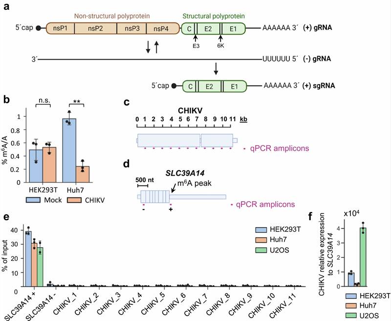
Figure 1 from Baquero-Pérez et al. (2024). Orthogonal validation of m6A modification status in viral RNA genomes. (a) LC-MS/MS m6A/A ratio quantification in poly(A)+ RNA from mock- and virus-infected cells. (b) Comparison of m6A-seq, SELECT, and nanopore direct RNA sequencing results for CHIKV RNA. (c) METTL3 depletion and its effect on viral replication. (d) Subcellular localization of m6A machinery components in infected cells. (e) Model: m6A-seq false positives in viral RNA arise from antibody cross-reactivity with structured RNA regions. (CC BY 4.0)
Representative m6A LC-MS Data Outputs
Our m6A LC-MS analysis pipeline delivers comprehensive multi-dimensional data outputs that provide a complete picture of m6A abundance and stoichiometry across experimental conditions. Below are representative examples of the key data types included in every project deliverable.
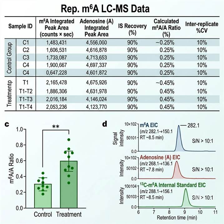
Representative m6A LC-MS data outputs. (Left) m6A quantification table with sample identifier, m6A and A integrated peak areas, isotope-labeled internal standard recovery (%), calculated m6A/A ratio (%), and inter-replicate %CV. (Center) Quantitative comparison of m6A/A ratios across control and experimental groups — individual data points displayed with group mean ± SD, statistical significance indicated. (Right) Overlaid extracted ion chromatograms (EICs) for m6A (m/z 282.1→150.1), adenosine (m/z 268.1→136.1), and 13C-m6A internal standard (m/z 288.1→156.1) showing baseline-resolved peaks with signal-to-noise ratios.
Every data deliverable includes raw chromatograms, calibration curves with linear regression parameters, QC performance metrics (accuracy, precision, LOD, LOQ), and a scientist consultation session for biological interpretation of m6A changes in the context of the epitranscriptome, RNA biology, and disease mechanisms. Custom reporting formats and integrated analysis with sequencing-based m6A mapping data are available on request.
Why Choose Our m6A LC-MS Analysis Services
Isotope Dilution Absolute Quantification
Our m6A quantification platform employs 13C-labeled m6A and 15N-labeled adenosine internal standards spiked at the earliest possible step, providing the highest accuracy for absolute m6A/A ratio measurements. Isotope dilution corrects for matrix effects, extraction efficiency variations, and ion suppression across diverse sample types, ensuring reliable cross-study comparability.
Multi-Platform Analytical Capability
We deploy both Orbitrap HRAM systems for broad-spectrum modified ribonucleoside discovery and triple quadrupole (QQQ) platforms in MRM mode for targeted high-sensitivity m6A quantification. This dual-platform configuration provides the analytical flexibility to match the specific requirements of each project — from unbiased RNA modification screening to trace-level m6A quantification.
Cross-Platform Integration Expertise
Our service bridges LC-MS quantification with sequencing-based m6A mapping approaches (MeRIP-seq, nanopore direct RNA-seq, SELECT), providing integrated multi-platform validation that combines the absolute quantification accuracy of mass spectrometry with the transcriptome-wide coverage of sequencing. This integrated approach is increasingly recognized as the gold standard for rigorous m6A research.
End-to-End Service and Expert Support
From experimental design consultation through sample processing, LC-MS acquisition, data analysis, and biological interpretation, our team of epitranscriptomics and mass spectrometry experts provides comprehensive support at every stage. Every project includes a dedicated scientist consultation session to ensure that m6A quantification data are interpreted in the correct biological context.
Related Services
Our m6A LC-MS analysis service is supported by a broader RNA modification and PTM characterization platform offering complementary analytical capabilities across modification types and research applications.
- Global PTM Profiling — Broad multi-PTM discovery analysis across diverse protein modification classes for integrated multi-omics studies
- MS-Based PTM Analysis — Comprehensive mass spectrometry platform for protein-level PTM discovery, quantification, and characterization
- Modified Peptide Enrichment Services — Specialized enrichment strategies for low-abundance modified peptides and nucleic acid modifications
- PTM Bioinformatics Analysis — Advanced bioinformatics for PTM data integration, functional annotation, and nucleic acid modification pathway analysis
- PTM Proteoform Mapping — Detailed characterization of combinatorial modification patterns on individual proteoforms
- Open-Search PTM Discovery — Unbiased open-search approach for detecting unexpected modifications across nucleic acids and proteins
- Bottom-Up MS-Based PTM Analysis — Deep PTM discovery and quantitative profiling using the shotgun proteomics approach
- PTM in Biological Research — Application-focused PTM analysis solutions for specific biological research contexts
- PTM Services — Comprehensive PTM analysis platform covering discovery, quantification, and characterization across all modification types
- PTM Quantification — Advanced quantitative PTM analysis services including absolute quantification, site occupancy, and multiplexed labeling approaches
Frequently Asked Questions
What is the m6A/A ratio and how is it calculated?
The m6A/A ratio represents the percentage of adenosine residues in an RNA sample that carry the N6-methyladenosine modification. It is calculated as: (peak area of m6A / peak area of adenosine) × 100, normalized to isotope-labeled internal standard recovery and calibrated against authentic m6A and A standards. Typical m6A/A ratios in mammalian mRNA range from 0.1% to 0.5%, depending on cell type, tissue, and biological condition.
What is the difference between LC-MS and MeRIP-seq for m6A analysis?
LC-MS provides direct, absolute quantification of global m6A/A ratios without antibody bias or amplification artifacts, delivering precise stoichiometric measurements across the entire RNA population. MeRIP-seq uses m6A-specific antibodies to enrich modified RNA fragments for sequencing, providing transcriptome-wide m6A distribution maps at single-base resolution but only relative enrichment levels (not absolute quantification). The two methods are complementary — LC-MS provides the quantitative accuracy and orthogonal validation essential for rigorous m6A research, while sequencing provides site-specific localization information.
What sample types and amounts are required for m6A LC-MS analysis?
For mRNA m6A quantification, we recommend ≥100 ng of poly(A)-selected mRNA (typically from ≥5 µg total RNA). For total RNA m6A analysis, ≥1 µg of purified total RNA is sufficient. For tRNA or small RNA analysis, ≥200 ng of purified small RNA is recommended. Our platform is compatible with RNA extracted from cultured cells, tissues (fresh-frozen or FFPE), biofluids, and viral particles. Smaller amounts may be acceptable for pilot studies or when sample is limited.
Can you distinguish m6A from m6Am and m1A?
Yes — our LC-MS platform can distinguish and independently quantify m6A, m6Am (N6,2′-O-dimethyladenosine), and m1A (N1-methyladenosine) in a single analytical run. While m6A and m1A share the same precursor and product ion masses (m6A/m1A are structural isomers), they are chromatographyically resolved by our optimized C18 gradient (m1A elutes earlier at ~6.1 min vs m6A at ~8.5 min). m6Am is distinguished from m6A by its distinct precursor mass (+14 Da from 2′-O-methylation). All three modifications are quantified using separate calibration curves with modification-specific internal standards.
How do you ensure the accuracy of m6A quantification?
Absolute quantification accuracy is ensured through multiple complementary measures: isotope dilution with 13C-m6A and 15N-A internal standards spiked at lysis to correct for recovery and matrix effects; external calibration with authentic m6A and A standards across 4 orders of dynamic range; system suitability standards, blank injections, and calibration verification at regular intervals; independent QC samples at low, medium, and high m6A/A levels in each batch; and inter-batch reproducibility monitoring through repeated analysis of pooled reference RNA samples.
Can LC-MS m6A quantification be integrated with sequencing-based approaches?
Yes — our integrated m6A validation package combines LC-MS absolute quantification with MeRIP-seq, nanopore direct RNA sequencing, or SELECT for cross-platform analysis. This integrated approach provides both the absolute quantification accuracy of LC-MS and the transcriptome-wide mapping resolution of sequencing, enabling rigorous validation of m6A sites and quantitative comparison across experimental conditions. We provide cross-platform correlation analysis, normalization between datasets, and unified reporting for integrated studies.
What are typical m6A levels in different RNA species?
Typical m6A/A ratios in mammalian cells range from 0.1% to 0.5% in mRNA (varying by cell type and condition), approximately 0.05–0.2% in total RNA (diluted by abundant rRNA which contains minimal m6A), and 0.01–0.1% in tRNA. m6Am is less abundant at approximately 0.01–0.05% of A in mRNA. These reference ranges are established from published LC-MS studies and validated against our in-house dataset across hundreds of samples. Significant deviations from these reference ranges may indicate dysregulation of m6A writer/eraser activity.
References
- Baquero-Pérez B, Yonchev ID, Delgado-Tejedor A, Medina R, Puig-Torrents M, Sudbery I, Begik O, Wilson SA, Novoa EM, Díez J. N6-methyladenosine modification is not a general trait of viral RNA genomes. Nat Commun. 2024;15:1964.
- Yang Y, Lu Y, Wang Y, Wen X, Qi C, Piao W, Jin H. Current progress in strategies to profile transcriptomic m6A modifications. Front Cell Dev Biol. 2024;12:1392159.
- Wang H, Hu X, Huang M, Liu J, Gu Y, Ma L, Zhou Q, Cao X. Mettl3-mediated mRNA m6A methylation promotes dendritic cell activation. Nat Commun. 2019;10:1898.
For research use only. Not for use in diagnostic procedures.