Arginine methylation has emerged as a focal point in biochemical research. This manuscript aims to elucidate the topic from the perspectives of background information, research techniques, methodologies, and results analysis.
What is Arginine Methylation?
Arginine methylation is a critical post-translational modification of proteins, involving the addition of methyl groups to the arginine residues of proteins by methyltransferases. This modification plays a pivotal role in various biological processes, including the regulation of gene expression, protein stability, signal transduction, and protein interactions within the nucleus and cytoplasm.
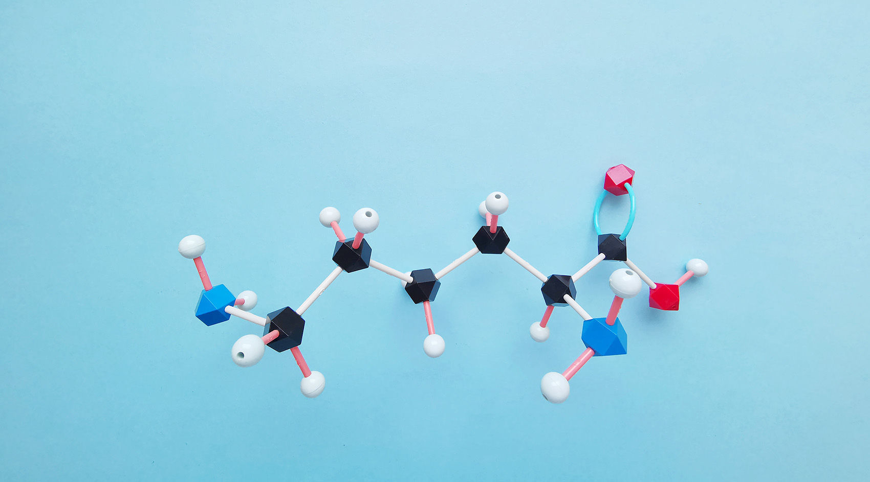
Arginine methylation primarily involves the transfer of a methyl group (CH₃) from S-adenosylmethionine (SAM) to the guanidino side chain of an arginine residue. This modification can manifest as monomethylation (a single methyl group), symmetric dimethylation (two methyl groups on the same nitrogen atom), or asymmetric dimethylation (two methyl groups on different nitrogen atoms).
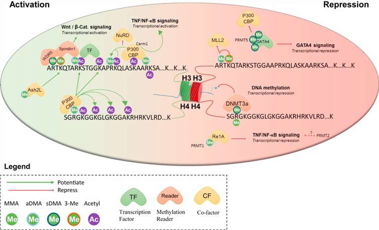 Figure 1. Arginine methylation in transcriptional regulation.
The catalytic process of arginine methylation is executed by specific enzymes known as protein arginine methyltransferases (PRMTs). These enzymes precisely recognize specific targets within proteins and catalyze the addition of methyl groups to arginine residues. Based on their catalytic characteristics, PRMTs can be classified into two types: Type I PRMTs primarily facilitate asymmetric dimethylation, while Type II PRMTs are responsible for symmetric dimethylation.
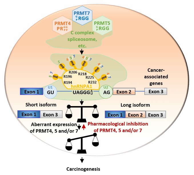 Figure 2. Working model of PRMT4, 5, and 7-mediated arginine methylation in alternative splicing regulation and cancer cell growth.
Biological Functions of Arginine Methylation
Regulation of Gene Expression
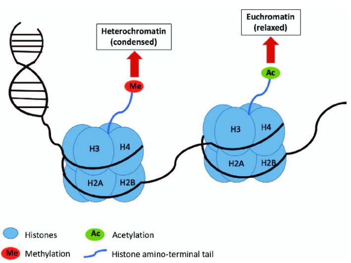
Methylation of arginine residues can significantly impact chromatin structure and gene transcription, primarily through modifications of histones and non-histone regulatory factors.
RNA Processing
Certain RNA-binding proteins, upon undergoing methylation, exhibit altered binding affinities with RNA. This modification influences various RNA-related processes, including mRNA splicing, transport, and stability.
Signal Transduction
Methylation of specific signaling proteins can modulate their activity or interactions with other proteins, thereby regulating signal transduction pathways.
Cell Cycle Regulation
Arginine methylation plays a crucial role in the regulation of key proteins involved in the cell cycle, thereby influencing cellular proliferation and differentiation.
Clinical Relevance
Aberrant arginine methylation is frequently associated with various pathological conditions, including cancer, cardiovascular diseases, and neurodegenerative disorders. Consequently, elucidating the precise biological mechanisms and regulatory networks of arginine methylation is of paramount importance for therapeutic interventions.
Research Methods for Arginine Methylation
The investigation of arginine methylation encompasses a variety of technical approaches and analytical metrics to elucidate its intricate biochemical pathways and regulatory mechanisms. The following are key methodologies and techniques employed in this research domain:
Mass Spectrometry
Mass spectrometry (MS) is a robust technique utilized for the identification and quantification of protein methylation. Samples are typically digested and subsequently analyzed using liquid chromatography-tandem mass spectrometry (LC-MS/MS) to accurately identify methylation sites and types. This method provides comprehensive information on the global methylation status of proteins.
Immunofluorescence
Immunofluorescence staining employs specific antibodies to label methylated arginine residues in cell or tissue sections, followed by observation using fluorescence microscopy. This technique allows for the visualization of the distribution and localization of methylated residues within cells, aiding in the understanding of their functional and dynamic changes.
Enzyme-Linked Immunosorbent Assay (ELISA)
ELISA utilizes methylation-specific antibodies to detect the methylation levels of specific proteins. This method is suitable for rapid screening and quantitative analysis, particularly when processing a large number of samples.
Chromatin Immunoprecipitation-Mass Spectrometry (ChIP-MS)
ChIP-MS combines chromatin immunoprecipitation (ChIP) with mass spectrometry to identify DNA and proteins associated with specific methylated arginine residues. This approach is particularly useful for studying how methylation influences protein-DNA interactions and gene expression.
Protein Microarrays
Protein microarray technology involves immobilizing a large number of proteins or peptides on a chip and using specific antibodies to detect their methylation status. This method enables the simultaneous analysis of the methylation states of multiple proteins, making it suitable for high-throughput analysis.
Results Analysis
Interpretation of Experimental Results on Arginine Methylation
To facilitate accurate comprehension and assessment of experimental results concerning arginine methylation, the following section elucidates two specific cases. These examples are intended to ensure that readers can accurately interpret and evaluate the outcomes of these experiments.
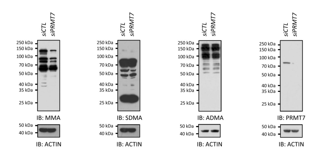 Figure 3. Analysis of Protein Methylation via Western Blot.
This figure illustrates the results of protein methylation analysis conducted using Western blotting. The image encompasses four distinct experimental outcomes, each pertaining to different types of methylated arginine residues:
Monomethylarginine (MMA)
Symmetric dimethylarginine (SDMA)
Asymmetric dimethylarginine (ADMA)
Specific protein arginine methyltransferase PRMT7
Each experiment presents results in two columns: control group (siCTL) and experimental group (siPRMT7). The control group exhibits normal expression, while the experimental group shows reduced expression of PRMT7. Detailed analysis is as follows:
Monomethylarginine (MMA)
Upon PRMT7 knockdown, the intensity of MMA-labeled protein bands appears diminished, suggesting that PRMT7 is involved in the regulation of monomethylation processes.
Symmetric Dimethylarginine (SDMA)
PRMT7 knockdown has minimal impact on SDMA methylation, with band intensities comparable to those in the control group.
Asymmetric Dimethylarginine (ADMA)
Similar to SDMA, PRMT7 knockdown does not significantly affect ADMA methylation, as band intensities remain consistent with those of the control group.
PRMT7 Protein
As anticipated, PRMT7 bands are markedly reduced or absent in the experimental group following PRMT7 knockdown.
Actin (ACTIN)
Actin serves as a loading control to ensure equal sample loading and accuracy of the protein transfer process. Actin bands exhibit stable expression across all experiments, thereby confirming the consistency and reliability of the experimental procedures.
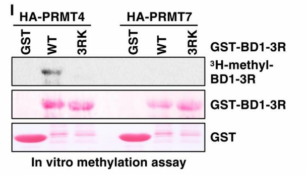 Figure 4. PRMT2 and PRMT4 interact and methylate BRD4.
This figure depicts an in vitro methylation assay comparing the methyltransferase activities of HA-PRMT4 and HA-PRMT7 on BRD4 protein. The experiment utilized wild-type (WT) and mutant (3RK) GST-BD1-3R BRD4 proteins as substrates.
The image displays three bands from top to bottom:
3H-methyl-BD1-3R: This row shows radiolabeled methylated BRD4. Significant signals are observed with PRMT4 and wild-type BRD4 (WT), indicating effective methylation by PRMT4. The signal disappears when using mutant BRD4 (3RK), suggesting these mutated residues are essential targets for PRMT4 methylation. In the PRMT7 columns, no significant methylation signals are observed for both wild-type and mutant BRD4, indicating weak or negligible methylation capability of PRMT7 under these in vitro conditions.
GST-BD1-3R: This row confirms the presence of BRD4 protein across all experimental samples through protein staining. Uniform protein bands are visible in all columns, indicating consistent protein loading.
GST: This row shows bands of GST protein as a control, confirming the presence and uniformity of GST protein across all samples.
Overall, this experiment demonstrates that PRMT4 specifically relies on certain arginine residues within BRD4 for methylation, which are disrupted in the 3RK mutant, abolishing methylation activity. In contrast, PRMT7 exhibits weak or no methylation activity towards BRD4 under these experimental conditions. These differences may arise from substrate specificity of the enzymes or variations in experimental conditions.
References
- Liu L, Lin B, Yin S, Ball LE, Delaney JR, Long DT, Gan W. Arginine methylation of BRD4 by PRMT2/4 governs transcription and DNA repair. Sci Adv. 2022
- Li WJ, He YH, Yang JJ, Hu GS, Lin YA, Ran T, Peng BL, Xie BL, Huang MF, Gao X, Huang HH, Zhu HH, Ye F, Liu W. Profiling PRMT methylome reveals roles of hnRNPA1 arginine methylation in RNA splicing and cell growth. Nat Commun. 2021
Our products and services are for research use only.

