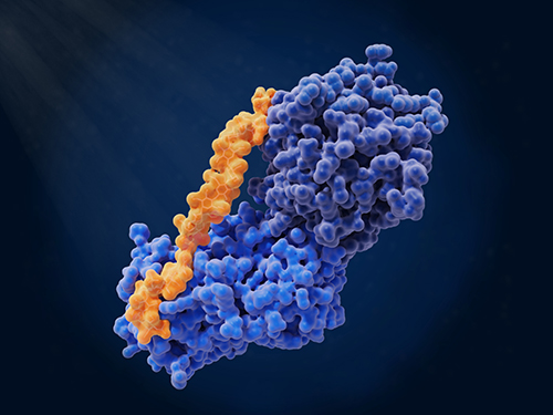
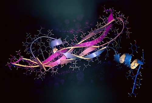
Protein crotonylation is a newly recognised type of protein modification that alters how genes are turned on or off. It works by attaching a crotonyl group (–CO–CH=CH₂) to lysine residues, changing the protein's charge and how it interacts with other molecules. This modification was first found on histones—the proteins that help package DNA—and is especially common in regions of DNA that are actively being used. Crotonylation is now known to influence key biological processes like cell development, sperm formation, and the suppression of tumour growth. Because it plays a role in both normal cell function and disease, scientists are using advanced proteomics tools to accurately detect and measure crotonylation across different proteins and conditions.
Biological Role and Disease Relevance
Histone crotonylation plays a key role in turning genes on by loosening the structure of chromatin, the protein-DNA complex that packages genetic material in the nucleus. This open chromatin state makes specific regions of DNA more accessible for transcription. Specialised proteins, known as "reader" proteins, recognise these crotonylation marks and help recruit other molecules that drive gene activation.
Importantly, crotonylation is not limited to histones. Researchers are increasingly studying crotonylation on non-histone proteins, where it appears to influence critical processes like cell signalling and immune responses. These functions suggest that crotonylation could be a broader regulatory mechanism than originally thought. In the context of cancer, abnormal patterns of crotonylation are linked to disrupted gene regulation, immune system avoidance, and tumour development.
Given its biological significance, analyzing crotonylation through advanced proteomics techniques offers a powerful approach to uncover how this modification contributes to disease. It also opens up new opportunities for identifying epigenetic biomarkers and therapeutic targets in cancer and other conditions.
Overview of Crotonylation Proteomics Workflow
The proteomics workflow for crotonylation analysis integrates biochemical, analytical, and computational steps to ensure high sensitivity, specificity, and reproducibility. The process typically includes:
- Sample preparation and protein extraction
- Enrichment of crotonylated peptides
- Mass spectrometry (MS) acquisition
- Bioinformatics analysis
- Functional validation
 Figure 1. Schematic diagram of the experimental procedures for mass spectrometry-based crotonylation analysis (Hou J Y, et al., 2021).
Sample Preparation and Protein Extraction
Accurate crotonylation analysis begins with optimised sample preparation to preserve labile crotonyl groups and ensure reliable downstream enrichment.
Sample Types and Collection
Crotonylation can be studied in a variety of biological samples, including:
- Cultured cell lines (e.g., HeLa, HEK293, cancer-derived lines)
- Fresh-frozen tissues or biopsies
- Formalin-fixed paraffin-embedded (FFPE) sections
Lysis Buffer Qptimisation
Cell or tissue lysis is performed using a high-salt or urea-based buffer containing:
- HDAC inhibitors (e.g., trichostatin A, sodium butyrate)
- Decrotonylase inhibitors (e.g., nicotinamide)
- Protease and phosphatase inhibitors (to maintain PTM integrity)
- Mechanical disruption (e.g., sonication or homogenization) is used to ensure complete cell lysis without heating the sample.
Protein Quantification and Digestion
After centrifugation to remove debris, supernatants are collected and protein concentration is measured using a BCA or Bradford assay. Samples are then:
- Reduced (e.g., with DTT) to break disulfide bonds
- Alkylated (e.g., with iodoacetamide) to prevent reformation
- Digested with trypsin at 37°C overnight to generate peptides.
- Optional: A second digestion with Lys-C can improve cleavage near crotonylated lysines.
Peptide Cleanup
Digested peptides are acidified and desalted using reversed-phase C18 columns to remove salts and inhibitors before enrichment.
Enrichment Techniques for Crotonylated Peptides
Crotonylated peptides typically comprise less than 1% of a complex digest, so targeted enrichment is essential for reliable detection. The gold-standard method is immunoaffinity purification using high-affinity anti-Kcr antibodies immobilised on agarose or magnetic beads. Key optimisations include:
- Pre-clearing with control beads or IgG to reduce nonspecific binding
- Antibody: peptide ratio titration to maximise capture without depleting low-abundance species
- Stringent washes to strip away unmodified peptides
- Acidic elution to release crotonylated peptides while preserving labile modifications
An orthogonal chemical approach employs hydroxylamine-based probes that react selectively with crotonyl lysines. After probe labelling, modified peptides are biotinylated and captured on streptavidin resin; subsequent elution and desalting yield an enriched fraction. Although chemical tagging can broaden coverage—especially for non-histone crotonylation—it requires additional cleanup and careful validation of reaction specificity.
For maximal depth, many labs combine both immunoaffinity and chemical-tag strategies in series or parallel, cross-validating sites and boosting sensitivity for low-stoichiometry crotonylated peptides.
Detection Methods for Crotonylation
Accurate detection of crotonylated peptides is essential for both the discovery and validation phases of proteomic studies. Several complementary techniques are used to achieve high sensitivity, specificity, and throughput:
Western Blotting (WB)
Western blotting is a widely adopted method for the qualitative detection of crotonylated proteins. Using pan-anti-crotonyllysine (Kcr) or site-specific antibodies enables rapid comparison of crotonylation levels across different experimental conditions, tissue types, or treatment groups. Though it does not provide site-level resolution or quantitative precision, WB is invaluable for:
- Validating LC-MS/MS results
- Screening for global crotonylation changes
- Assessing antibody specificity
Advantages include ease of use, low cost, and fast turnaround time, making WB an ideal preliminary tool during early-stage analysis or functional follow-up experiments.
Protein Microarrays
Protein microarrays allow for high-throughput detection of crotonylation across hundreds to thousands of proteins in parallel. In this approach, target proteins or peptides are immobilised on array surfaces and probed with anti-Kcr antibodies. Detection is typically performed via fluorescence- or chemiluminescence-based readouts.
Applications of protein microarrays include:
- Comparative crotonylation profiling across cell lines or patient samples
- Identification of potential crotonylation substrates
- Drug screening studies targeting epigenetic modifiers
While less sensitive than MS-based approaches, microarrays excel in speed, scalability, and compatibility with large sample sets, making them suitable for exploratory or screening-focused studies.
Liquid Chromatography-Tandem Mass Spectrometry (LC-MS/MS)
High-resolution LC-MS/MS remains the gold standard for site-specific identification and quantification of crotonylated peptides. Following antibody-based enrichment of crotonylated peptides, samples are subjected to a multi-step analytical workflow:
- NanoLC Separation:
Peptides are separated using nano-scale reverse-phase liquid chromatography to reduce complexity and increase dynamic range.
- Electrospray Ionization (ESI):
Separated peptides are ionized into the gas phase using ESI, enabling their detection by the mass spectrometer.
- High-Resolution MS Analysis:
Instruments such as the Orbitrap Fusion Lumos, Q Exactive, or Bruker timsTOF Pro acquire MS¹ scans with high resolution and mass accuracy, essential for distinguishing crotonylated species from other modifications.
- Peptide Fragmentation:
HCD (Higher-energy Collisional Dissociation): Generates b/y-ion series suitable for sequencing and confident site localisation.
ETD (Electron Transfer Dissociation): Preserves labile PTMs like crotonylation, particularly beneficial for heavily modified or histone-derived peptides.
- Targeted Quantification (Optional):
For targeted validation, Parallel Reaction Monitoring (PRM) or Selected Reaction Monitoring (SRM) can be employed to reproducibly quantify specific crotonylation sites across multiple samples.
Data Processing and Quantitative Analysis
Accurate data processing is essential for identifying crotonylation sites and quantifying their abundance. After LC-MS/MS acquisition, raw data files are processed using specialised software such as MaxQuant, Proteome Discoverer, or PEAKS Studio, which support post-translational modification searches.
Database Searching and PTM Identification
- The software compares MS/MS spectra against a reference protein database (e.g., UniProt).
- Crotonylation (Kcr) is set as a variable modification on lysine residues, with a mass shift of +68.0262 Da.
- False discovery rate (FDR) is typically controlled at 1% at both the peptide and protein levels using a decoy database strategy.
- High-confidence site localisation is ensured using site probability scores.
Quantitative Strategies
- labell-Free Quantification (LFQ): Measures peptide ion intensities across runs for relative quantitation.
- Isobaric Tagging (TMT/iTRAQ): Enables multiplexed comparison of multiple conditions in a single run.
- Normalization techniques (e.g., total ion current, median centering) are applied to correct for technical variation.
Data Filtering and Statistical Analysis
- Only peptides with confidently assigned crotonylation sites and consistent replicate detection are retained.
- Fold change and p-values are calculated to identify significantly regulated crotonylation events.
- Volcano plots and hierarchical clustering can visualize PTM dynamics across experimental conditions.
Functional Annotation
- Identified proteins are annotated using tools like DAVID, GO, or KEGG to uncover enriched biological processes and pathways.
- PTM crosstalk with acetylation or methylation can also be explored using integrated datasets.
Validation and Functional Characterization
Following the identification of crotonylation sites by mass spectrometry, rigorous validation and functional analysis are essential to confirm site specificity and determine biological relevance.
Experimental Validation Approaches
- Western Blotting with Anti-Kcr Antibodies: Confirms the presence of crotonylation at the protein level. Site-specific antibodies, when available, provide higher confidence than pan-Kcr reagents.
- Targeted MS (PRM/SRM): PRM or SRM enables precise quantification of specific crotonylated peptides across multiple samples, enhancing reproducibility.
- Site-Directed Mutagenesis: Substituting lysine residues (e.g., K→R or K→Q) allows researchers to assess whether crotonylation at specific sites affects protein function, stability, or localisation.
Functional Characterization Strategies
- Chromatin Immunoprecipitation (ChIP): Used to map crotonylation-enriched regions on chromatin, especially in histones, and correlate with gene expression activity.
- Reporter Gene Assays: Evaluate how crotonylated histones or transcription factors influence promoter activity and gene expression.
- Protein–Protein Interaction Analysis: Co-immunoprecipitation (Co-IP), BioID, or proximity labelling can reveal how crotonylation modulates protein complexes and interaction networks.
- Cellular Phenotypic Assays: Assess downstream biological effects such as cell proliferation, differentiation, or apoptosis in response to altered crotonylation states.
Select Service
Applications in Cancer and Drug Discovery
Crotonylation has emerged as a key epigenetic marker with significant implications in cancer biology and therapeutic innovation. Its dynamic regulation by metabolic state and chromatin-associated enzymes positions it as both a functional readout and a potential intervention point in cancer.
Cancer Biomarker Discovery
Quantitative proteomics of crotonylation enables the identification of cancer-specific signatures. For example, decreased crotonylation at H3K18 is observed in liver and colorectal cancers, correlating with poor prognosis. Such site-specific crotonylation patterns offer promise as diagnostic or prognostic biomarkers, especially when combined with histological or multi-omics data.
Epigenetic Drug Response Monitoring
HDAC inhibitors, commonly used in epigenetic therapy, modulate global crotonylation levels. Proteomic analysis can monitor therapeutic response by quantifying changes in histone and non-histone crotonylation, helping to identify sensitive pathways and resistance mechanisms.
Target Validation and Mechanistic Insights
Crotonylation affects the function of transcription factors, coactivators, and metabolic enzymes. Functional proteomics can validate targets by linking crotonylation to altered protein activity, gene regulation, or tumour cell behavior. For instance, crotonylation of p53 may alter its DNA binding affinity, influencing apoptosis and cell cycle control.
Immuno-oncology Applications
Crotonylation has been implicated in immune evasion and inflammation. Mapping crotonylation in tumour-associated macrophages or T cells can reveal epigenetic reprogramming linked to immune suppression. This supports drug development aimed at reactivating anti-tumour immunity through epigenetic remodeling.
Companion Diagnostics Development
Integrating crotonylation profiling with clinical genomics can inform personalized therapy. tumours with specific crotonylation-dependent gene expression profiles may benefit from tailored combinations of metabolic modulators and epigenetic drugs.
Related Article
Challenges and Considerations
Antibody Specificity and Batch Variability: The success of immunoaffinity enrichment depends heavily on the quality of anti-crotonyl lysine (Kcr) antibodies. Non-specific binding or cross-reactivity with structurally similar PTMs (e.g., acetylation) can lead to false-positive identifications. Batch-to-batch variation further complicates reproducibility across studies.
Low Abundance of Crotonylated Peptides: Crotonylation is typically a low-abundance modification, especially in non-histone proteins. Without efficient enrichment and sensitive MS settings, crotonylated peptides may go undetected, reducing coverage and limiting biological interpretation.
False Site Assignments: Incomplete fragmentation during MS or mislocalisation of modification sites by search algorithms can produce inaccurate results. This is particularly problematic for lysine-rich sequences, where multiple potential modification sites exist in close proximity.
Sample Integrity and PTM Stability: Crotonyl groups are chemically labile and susceptible to loss during sample preparation if deacetylase inhibitors are not used. Even minor delays or suboptimal buffer conditions can significantly alter modification profiles.
Data Interpretation Complexity: Differentiating biologically meaningful changes from technical noise requires rigorous statistical analysis and biological replication. Moreover, the functional impact of most crotonylation sites remains poorly characterized, making downstream interpretation more speculative without validation.
Case study
Case: Quantitative Crotonylome Analysis Expands the Roles of p300 in the Regulation of Lysine Crotonylation Pathway
Background
Lysine crotonylation (Kcr) is a post-translational modification (PTM) involved in gene regulation. While p300 is known to catalyze histone crotonylation, its role in non-histone protein crotonylation remains unclear.
Purpose
To investigate how p300 regulates the global crotonylome and to identify crotonylated substrates and biological processes affected by p300-mediated crotonylation.
Method
- Used SILAC-based quantitative proteomics in wild-type and p300-knockout HCT116 cells.
- Pan-Kcr antibody enrichment followed by LC-MS/MS analysis.
- Employed bioinformatic tools (GO, Reactome, STRING) for functional and pathway analysis.
Results
- Identified 816 Kcr sites, 775 of which were quantified.
- p300 deletion led to downregulation of 88 Kcr sites and upregulation of 31 sites.
- Affected proteins are involved in RNA metabolism, translation, and chromatin remodeling.
- Several p300-regulated Kcr proteins are linked to cancer pathways and protein–protein interaction networks.
 Figure 2. Systematic Profiling of Kcr Proteome.
Conclusion
p300 regulates a broad set of non-histone Kcr sites, expanding its role in gene expression and disease processes. This study offers the first quantitative crotonylome landscape dependent on p300 and provides a foundation for exploring Kcr functions in health and disease.
Relevant FAQ
Why is enrichment necessary before MS analysis?
Crotonylated peptides are often low in abundance and can be masked by unmodified peptides. Enrichment using anti-Kcr antibodies isolates crotonylated peptides, enhancing MS detection sensitivity and site coverage.
What MS platforms are recommended for crotonylation studies?
High-resolution instruments like Orbitrap Fusion, Q Exactive, and timsTOF Pro are preferred for their sensitivity, resolution, and ability to confidently localise modification sites.
Can crotonylation be quantified across multiple conditions or samples?
Yes. Quantitative strategies such as LFQ, iTRAQ/TMT, or SILAC can be used to compare crotonylation levels across experimental groups or treatment conditions.
What downstream applications can benefit from crotonylation analysis?
Applications include biomarker discovery, epigenetic drug development, functional genomics, and mechanistic studies of cancer and metabolic regulation.
References
- Hou J Y, et al. Emerging roles of non-histone protein crotonylation in biomedicine. Cell & Bioscience, 2021, 11(1): 101. DOI: 10.1186/s13578-021-00616-2
- Wei W, et al. Large-scale identification of protein crotonylation reveals its role in multiple cellular functions. Journal of proteome research, 2017, 16(4): 1743-1752. DOI: 10.1021/acs.jproteome.7b00012
- Jiang G, Li C, Lu M, et al. Protein lysine crotonylation: past, present, perspective. Cell death & disease, 2021, 12(7): 703. DOI: 10.1038/s41419-021-03987-z
Our products and services are for research use only.


