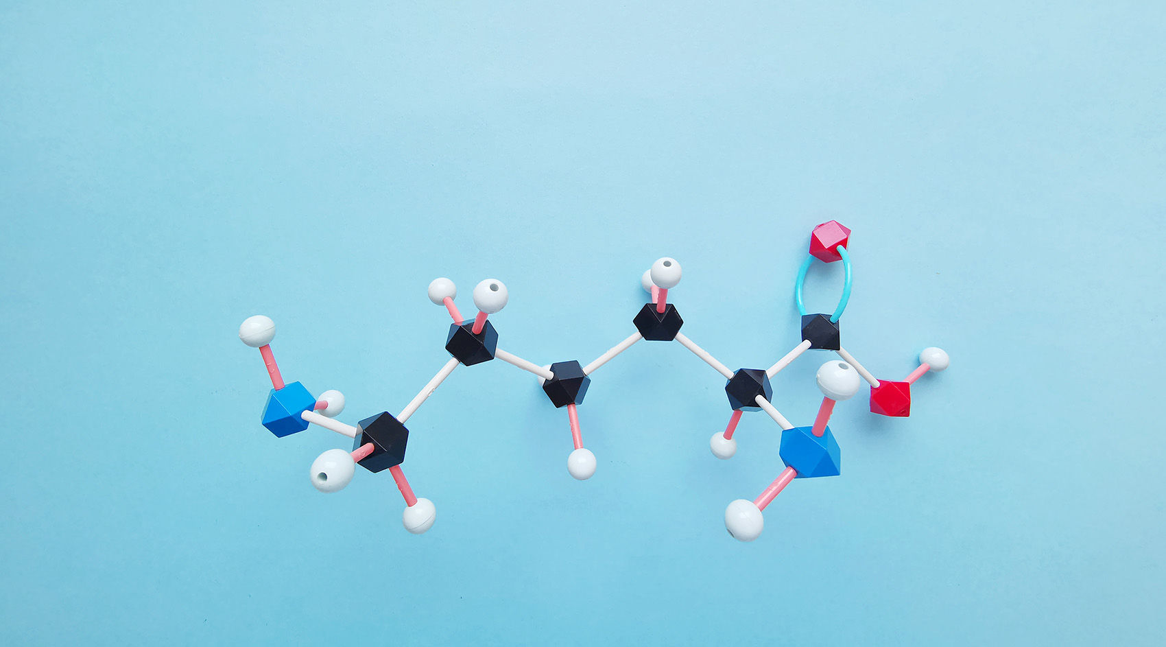
Protein crotonylation is a form of lysine acylation first spotted on histones in 2011 using high-resolution mass spectrometry. In this process, a crotonyl group (–CO–CH=CH–CH₃) is transferred from crotonyl-CoA onto the side chain of lysine amino acids. Compared to the smaller, more flexible acetyl group, the crotonyl moiety is bulkier, more water-repellent, and contains a flat double bond—features that give it unique chemical behavior and influence how proteins fold and interact.
Early studies found crotonylation enriched in sperm and egg cells, where it sits on "open" chromatin regions to promote gene activity. Unlike acetylation, which often overlaps in function with other marks, crotonylation appears to switch on very specific sets of genes, especially those involved in development, metabolism, and cell fate decisions. Its precise placement and removal by specialized enzymes help cells fine-tune their genetic programs in response to changing conditions.
Select Service
Biological Significance and Functional Roles
Protein crotonylation is a key molecular signal that helps control how genes are turned on or off, how DNA is packaged in the cell, and how proteins function. It was first discovered on histones—the proteins that wrap DNA—where crotonyl groups are added to specific lysine sites like H3K18 and H3K9. These modified sites are often found at the start of active genes and in regions that boost gene expression, such as enhancers. Compared to acetylation, crotonylation is more tightly linked to long-term gene activity, especially during critical stages of development like sperm formation.
But crotonylation does not stop at histones. It also occurs on many other proteins involved in essential cell processes like metabolism, signal transmission, and stress adaptation. In these non-histone proteins, crotonylation can influence how stable the protein is, how well it works, and where it goes inside the cell. This adds a versatile layer of control that helps maintain balance in the cell's internal environment.
In disease, abnormal patterns of protein crotonylation have been linked to cancer growth, chronic inflammation, and metabolic dysfunction. In liver and colorectal cancers, changes in histone crotonylation have been shown to affect the activity of genes that drive tumour development and uncontrolled cell growth. These disruptions suggest that crotonylation plays a key role in how cancer cells behave. Because of this, crotonylation is gaining attention as both a potential disease marker and a target for new epigenetic drugs designed to modify gene activity without altering the DNA sequence itself.
Molecular Basis and Mechanism of Crotonylation
Protein crotonylation is a lysine acylation modification in which a crotonyl group (C₄H₅O) is covalently attached to the ε-amino group of lysine residues. This process is catalysed by crotonyltransferases using crotonyl-CoA as the donor substrate. The reverse reaction—removal of the crotonyl group—is mediated by decrotonylases, which restore the unmodified lysine residue.
Crotonylation Writers: Crotonyltransferases
The primary enzymes known to catalyse crotonylation are p300/CBP, well-characterized histone acetyltransferases that also exhibit crotonyltransferase activity. These enzymes transfer the crotonyl group from crotonyl-CoA to target lysine residues on histones and non-histone proteins. Crotonyl-CoA itself is generated through cellular metabolism, particularly from fatty acid oxidation and short-chain fatty acid catabolism. Thus, crotonylation levels are tightly linked to metabolic state.
Crotonylation Erasers: Decrotonylases
Crotonyl marks are removed by class I histone deacetylases (HDAC1, HDAC2, and HDAC3) and members of the sirtuin family, notably SIRT1, SIRT2, and SIRT3. These NAD⁺-dependent enzymes recognise crotonylated lysines and catalyse hydrolytic cleavage of the crotonyl group. The activity of these enzymes is modulated by cellular redox and energy status, providing a mechanism by which metabolic fluctuations influence chromatin structure and gene expression.
Substrate Specificity and Selectivity
Not all lysine residues are equally modified. Crotonylation often occurs at functionally important residues within transcriptionally active chromatin regions, such as histone H3K18 and H4K8. Substrate preference is influenced by chromatin accessibility, local sequence motifs, and cofactor availability.
 Figure 1. The modulation of protein crotonylation.
Key Enzymes and Regulatory Proteins
Protein crotonylation is dynamically regulated by three main classes of proteins: writers, erasers, and readers, which control the addition, removal, and interpretation of crotonyl marks, respectively.
- Readers: YEATS domain–containing proteins (e.g., AF9, ENL) bind crotonyl-modified histones with high affinity, recruiting transcriptional coactivators.
- Writers: Novel KCTs outside of p300/CBP are under investigation for non-histone substrates.
- Erasers: Class I HDACs (HDAC1/2) display broader deacylase activity, whereas SIRT family members exhibit substrate and context specificity.
Crosstalk with Other Epigenetic Marks
Protein crotonylation does not act in isolation but integrates into the wider "histone code," where multiple post-translational modifications converge to regulate chromatin structure and gene expression. Key aspects include:
Mutual Exclusivity and Competition
Crotonylation and acetylation both target ε-amino groups of lysine residues. At sites such as H3K18 and H4K16, crotonylation can outcompete acetylation, limiting deacetylase (HDAC) binding and promoting a more open chromatin conformation that favors transcriptional activation.
Sequential Modification
In some promoters, initial acetylation by p300/CBP creates a permissive chromatin environment that enhances subsequent crotonylation, amplifying transcriptional responses to stimuli. Conversely, removal of crotonyl marks by SIRT3 may facilitate downstream methylation, enabling a switch from active to repressive chromatin states.
Reader Protein Specificity
YEATS domain–containing "reader" proteins distinguish crotonyl from acetyl marks—AF9 and ENL bind crotonyl-modified histones with greater affinity, recruiting the super-elongation complex to drive RNA polymerase II elongation. This selective recognition underscores how crotonylation can uniquely influence gene programs.
Integration with Ubiquitination and Methylation
At enhancers, H2BK120 ubiquitination can enhance local crotonyl-CoA availability, promoting nearby histone crotonylation. Simultaneously, crosstalk with H3K4 methylation coordinates the recruitment of transcription factors, illustrating a multilayered regulatory network.
Analytical Techniques for Crotonylation Detection
Accurate detection of crotonylation sites is essential for understanding their biological function. Due to the typically low abundance and dynamic nature of crotonylation, specialized enrichment and high-sensitivity analytical methods are required. The following techniques represent the current gold standard in crotonylation analysis:
Mass Spectrometry–Based Proteomics
Mass spectrometry (MS) is the most powerful technique for site-specific identification and quantification of crotonylated peptides.
- Sample Preparation: Proteins are digested into peptides (commonly with trypsin), and crotonylated peptides are enriched using immunoprecipitation before MS analysis.
- Instrumentation: High-resolution MS systems, such as Orbitrap or Q-TOF platforms, allow detection of crotonylation-specific mass shifts (+68.0266 Da on lysine residues).
- Fragmentation Methods: Higher-energy collisional dissociation (HCD) or electron transfer dissociation (ETD) are used to preserve PTM information during peptide fragmentation.
Antibody-Based Enrichment
Due to the low stoichiometry of crotonylation, enrichment is essential before MS.
- Pan-Kcr Antibodies: These antibodies recognise a broad range of crotonyl-lysine sites and are used for global crotonylome profiling.
- Site-Specific Antibodies: Designed to target specific crotonylated lysine residues (e.g., H3K18cr), enhancing site-level analysis and biological interpretation.
- Cross-reactivity Considerations: Antibody validation is critical to avoid overlap with structurally similar acylations like acetylation or butyrylation.
Quantitative Analysis Strategies
Quantitative assessment enables comparison of crotonylation levels across conditions.
- Label-Free Quantification (LFQ): Relies on ion intensity comparison across LC-MS runs. Suitable for large sample sets but sensitive to run-to-run variation.
- Isobaric Tagging (TMT/iTRAQ): Allows multiplexing of samples for relative quantification with reduced technical variability.
- Metabolic Labeling (SILAC): Integrates heavy isotopes during cell culture, enabling highly accurate quantification in controlled systems.
Bioinformatics and Data Interpretation
Following data acquisition, specialized software is used to process crotonylation data:
- Search Engines: Tools like MaxQuant, Proteome Discoverer, and pFind can detect crotonylated peptides by incorporating specific mass shifts in search parameters.
- Site Localization: Probabilistic scoring (e.g., Andromeda, Ascore) assigns crotonylation to specific lysine residues.
- Functional Annotation: Crotonylated proteins are mapped to cellular pathways, disease associations, and protein interaction networks.
Quantitative Crotonylation Profiling: Label-Free vs. Label-Based Approaches
| Method | Description | Advantages | Limitations | Best Suited For |
|---|---|---|---|---|
| Label-Free Quantification | Measures peptide ion intensities across runs using high-resolution MS. | -Cost-effective -Unlimited sample number -Simple workflow |
- Sensitive to run-to-run variation - Requires strong alignment algorithms |
Comparative profiling in large cohorts |
| TMT/iTRAQ | Tags peptides with mass reporter ions for multiplexed MS analysis. | -High multiplexing -Reduced technical variation |
- Ratio compression - Requires MS3 for accuracy |
High-throughput, multi-sample experiments |
| SILAC | Incorporates heavy amino acids into proteins during cell growth. | -Accurate quantification -Low technical variability |
- Limited to cell culture - Not applicable to tissues |
Dynamic studies in live cells |
| DIA | Fragmentation of all ions within a range for reproducible quantification. | -High reproducibility -Broad proteome coverage |
- Complex data analysis - Requires spectral library |
Global crotonylation profiling |
Select Service
Related Article
Applications in Disease Research and Drug Discovery
Cancer Biology
Non-histone crotonylation in metastasis: In hepatocellular carcinoma (HCC), site-specific crotonylation of the cytoskeletal GTPase SEPT2 at lysine 74 enhances cell invasiveness via the SEPT2-K74cr → P85α → AKT axis. SEPT2-K74cr levels are significantly higher in tumours from patients with early recurrence, and mutation of K74 to a non-crotonylatable residue reduces metastasis in vitro and in vivo (Zhang X, et al., 2023).
Hypoxia-induced lamin A crotonylation: Under tumour hypoxia, lamin A becomes hyper-crotonylated at K265/270, promoting proliferation and preventing senescence. Quantitative TMT MS profiling of 12 paired HCC and adjacent tissues identified >3,700 crotonylation sites, and HDAC6 was shown to act as the decrotonylase for lamin A, suggesting HDAC6 inhibitors could elevate lamin A K265/270cr to modulate growth (Zhang D, et al., 2022).
 Figure 2. Graphic model showing the effect of SEPT2-K74 crotonylation in HCC (Zhang X, et al., 2023).
Metabolic and Inflammatory Diseases
MASLD and mitochondrial-ER contact: In patients with metabolic dysfunction–associated steatotic liver disease (MASLD), global protein crotonylation—and specifically ALR (K78) crotonylation—are markedly reduced. Loss of ALR K78cr impairs ALR–MFN2 interaction, disrupts mitochondria-ER contacts, and exacerbates lipid accumulation, highlighting crotonylation restoration as a novel therapeutic angle (Wang X, et al., 2025).
Immune regulation: Although detailed studies are emerging, crotonyl-CoA fluctuations in activated macrophages suggest crotonylation writers and erasers could be targeted to fine-tune inflammatory gene expression.
 Figure 3. Graphic of Lysine crotonylation of ALR in deterioration of hepatic steatosis (Wang X, et al., 2025).
Challenges and Future Directions in Crotonylation Research
Low Abundance and Stoichiometry
Crotonylation is a low-abundance PTM, often present at substoichiometric levels compared to acetylation. This limits detection sensitivity, particularly in complex biological samples such as tissues or plasma.
Limited Site-Specific Antibodies
Most commercially available antibodies recognise crotonyl-lysine in general but lack site specificity. This hampers the precise validation of individual modification sites and their biological relevance.
Incomplete Enzyme Characterization
The full range of enzymes responsible for writing, erasing, and reading crotonylation—especially on non-histone proteins—remains poorly defined. This knowledge gap restricts mechanistic studies and drug target development.
Lack of Functional Annotations
Most identified crotonylation sites are cataloged without known functions. Functional validation through mutagenesis, structural analysis, or interaction studies is still limited and labor-intensive.
Multi-PTM Crosstalk Complexity
Crotonylation coexists with other lysine modifications, such as acetylation, succinylation, and ubiquitination. Dissecting this PTM crosstalk at specific residues is technically challenging due to overlapping mass shifts and antibody cross-reactivity.
Relevant FAQ
How is crotonylation different from acetylation?
While both are lysine acylations, crotonylation adds a bulkier, planar group that can create distinct structural and functional effects compared to acetylation.
What types of samples are suitable for crotonylation profiling?
Both cultured cells and clinical tissue samples can be analysed, provided high-quality protein extraction and enrichment protocols are used.
How do metabolic changes influence crotonylation levels?
Crotonylation levels are sensitive to intracellular crotonyl-CoA, which is regulated by fatty acid oxidation and short-chain fatty acid metabolism, linking metabolism to epigenetic regulation.
What is the significance of crotonylation in stem cells and development?
Crotonylation marks active enhancers and promoters in pluripotent stem cells, and changes in crotonylation profiles are associated with cell fate decisions and differentiation.
What are the main challenges in crotonylation research?
Challenges include low stoichiometry, lack of site-specific antibodies, limited known substrates, and difficulty in dynamic quantification.
References
- Jiang G, Li C, Lu M, et al. Protein lysine crotonylation: past, present, perspective. Cell death & disease, 2021, 12(7): 703. DOI: 10.1038/s41419-021-03987-z
- Wang S, Mu G, Qiu B, et al. The function and related diseases of protein crotonylation. International Journal of Biological Sciences, 2021, 17(13): 3441. DOI: 10.7150/ijbs.58872
- Xu W, Wan J, Zhan J, et al. Global profiling of crotonylation on non-histone proteins. Cell research, 2017, 27(7): 946-949. DOI: 10.1038/cr.2017.60
Our products and services are for research use only.


